Anti-PDE7A抗体
产品名称: Anti-PDE7A抗体
英文名称: PDE7A
产品编号: YB--11575R
产品价格: null
产品产地: 中国/美国
品牌商标: Ybscience
更新时间: 2023-08-17T10:29:50
使用范围: 科研使用
上海钰博生物科技有限公司
- 联系人 : 陈环环
- 地址 : 上海市沪闵路6088号龙之梦大厦8楼806室
- 邮编 : 200612
- 所在区域 : 上海
- 电话 : 183****2235
- 传真 : 021-60514606
- 邮箱 : shybio@126.com
Anti-PDE7A抗体
| 产品编号 | YB-11575R |
| 英文名称 | PDE7A |
| 中文名称 | 磷酸二酯酶7抗体 |
| 别 名 | HCP 1; HCP1; High affinity cAMP specific 3'5' cyclic phosphodiesterase 7A; High affinity cAMP-specific 3''; P2A; PDE 7; PDE 7A; PDE7; PDE71; PDE7A; PDE7A_HUMAN; Phosphodiesterase 7A; Phosphodiesterase isozyme 7; Phosphodiesterase7A; Rolipram insensitive phosphodiesterase type 7; TM 22; TM22; 5''-cyclic phosphodiesterase 7A. |
| 规格价格 | 100ul/1380元 购买 200ul/2200元 购买 大包装/询价 |
| 说 明 书 | 100ul 200ul |
| 研究领域 | 心血管 细胞生物 神经生物学 信号转导 激酶和磷酸酶 |
| 抗体来源 | Rabbit |
| 克隆类型 | Polyclonal |
| 交叉反应 | Human, Mouse, Rat, Chicken, Dog, Pig, Cow, Horse, Rabbit, |
| 产品应用 | WB=1:500-2000 ELISA=1:500-1000 IHC-P=1:400-800 IHC-F=1:400-800 Flow-Cyt=1μg/Test ICC=1:100-500 IF=1:100-500 (石蜡切片需做抗原修复) not yet tested in other applications. optimal dilutions/concentrations should be determined by the end user. |
| 分 子 量 | 55kDa |
| 细胞定位 | 细胞浆 |
| 性 状 | Lyophilized or Liquid |
| 浓 度 | 1mg/ml |
| 免 疫 原 | KLH conjugated synthetic peptide derived from human PDE7A (341-440aa):341-440/482 |
| 亚 型 | IgG |
| 纯化方法 | affinity purified by Protein A |
| 储 存 液 | 0.01M TBS(pH7.4) with 1% BSA, 0.03% Proclin300 and 50% Glycerol. |
| 保存条件 | Store at -20 °C for one year. Avoid repeated freeze/thaw cycles. The lyophilized antibody is stable at room temperature for at least one month and for greater than a year when kept at -20°C. When reconstituted in sterile pH 7.4 0.01M PBS or diluent of antibody the antibody is stable for at least two weeks at 2-4 °C. |
| PubMed | PubMed |
| 产品介绍 | background: Phosphodiesterases (PDE, also designated cyclic nucleotide phosphodiesterase) are important for the downregulation of the intracellular level of the second messenger cyclic adenosine monophosphate (cAMP) by hydrolyzing cAMP to 5'AMP. Phosphodiesterase type 3 isoforms, PDE3A and 3B, are expressed primarily in cardiovascular tissue and adipose tissue, respectively. PDE3A, is found in myocardium and platelets and PDE3B is found in lymphocytes. The PDE7A1 (HCP1) isozyme and the PDE7A2 proteins, alternate splice products of PDE7A, are highly expressed in skeletal muscle. PDE7B is most highly expressed in pancreas. The PDE family contains proteins that serve tissue-specific roles in regulation of lipolysis, glycogenolysis, myocardial contractility, and smooth muscle relaxation. Function: Hydrolyzes the second messenger cAMP, which is a key regulator of many important physiological processes. May have a role in muscle signal transduction. Subunit: Interacts with CBFA2T3. Subcellular Location: Cytoplasm Tissue Specificity: PDE7A1 is found at high levels in skeletal muscle and at low levels in a variety of tissues including brain and heart. It is expressed as well in two T-cell lines. PDE7A2 is found abundantly in skeletal muscle and at low levels in heart. Similarity: Belongs to the cyclic nucleotide phosphodiesterase family. PDE7 subfamily. SWISS: Q13946 Gene ID: 5150 Database links: Entrez Gene: 5150 Human Omim: 171885 Human SwissProt: Q13946 Human Unigene: 527119 Human Unigene: 728847 Human Important Note: This product as supplied is intended for research use only, not for use in human, therapeutic or diagnostic applications. |
| 产品图片 |
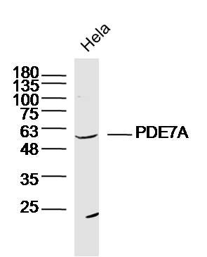 Sample: Hela Cell (Human) Lysate at 30 ug
Primary: Anti-PDE7A (bs-11575R) at 1/300 dilution Secondary: IRDye800CW Goat Anti-Rabbit IgG at 1/20000 dilution Predicted band size: 55kD Observed band size: 55kD  Sample: Muscle (Mouse)Lysate at 40 ug
Primary: Anti-PDE7A(bs-11575R)at 1/300 dilution Secondary: IRDye800CW Goat Anti-RabbitIgG at 1/20000 dilution Predicted band size: 55kD Observed band size: 55kD  Sample:Brain (Mouse)Lysate at 40 ug
Primary: Anti-PDE7A(bs-11575R)at 1/300 dilution Secondary: IRDye800CW Goat Anti-RabbitIgG at 1/20000 dilution Predicted band size: 55kD Observed band size: 55kD 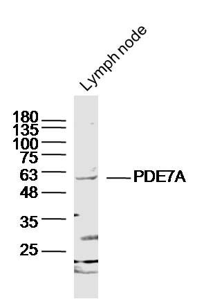 Sample: Lymph node (Mouse) Lysate at 40 ug
Primary: Anti-PDE7A (bs-11575R) at 1/300 dilution Secondary: IRDye800CW Goat Anti-Rabbit IgG at 1/20000 dilution Predicted band size: 55kD Observed band size: 55kD 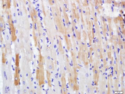 Paraformaldehyde-fixed, paraffin embedded (rat heart); Antigen retrieval by boiling in sodium citrate buffer (pH6.0) for 15min; Block endogenous peroxidase by 3% hydrogen peroxide for 20 minutes; Blocking buffer (normal goat serum) at 37°C for 30min; Antibody incubation with (PDE7A) Polyclonal Antibody, Unconjugated (bs-11575R) at 1:500 overnight at 4°C, followed by a conjugated secondary (sp-0023) for 20 minutes and DAB staining.
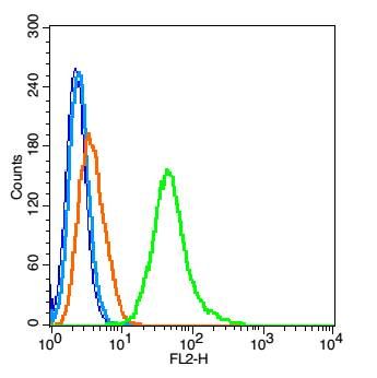 Blank control(blue): RSC96 cells(fixed with 2% paraformaldehyde (10 min) , then permeabilized with 90% ice-cold methanol for 30 min on ice).
Primary Antibody:Rabbit Anti- PDE7A antibody(bs-11575R), Dilution: 1μg in 100 μL 1X PBS containing 0.5% BSA; Isotype Control Antibody: Rabbit IgG(orange) ,used under the same conditions ); Secondary Antibody: Goat anti-rabbit IgG-PE(white blue), Dilution: 1:200 in 1 X PBS containing 0.5% BSA. |