Anti-Optineurin抗体
产品名称: Anti-Optineurin抗体
英文名称: Optineurin
产品编号: YB--13658R
产品价格: null
产品产地: 中国/美国
品牌商标: Ybscience
更新时间: 2023-08-17T10:29:50
使用范围: 科研使用
上海钰博生物科技有限公司
- 联系人 : 陈环环
- 地址 : 上海市沪闵路6088号龙之梦大厦8楼806室
- 邮编 : 200612
- 所在区域 : 上海
- 电话 : 183****2235
- 传真 : 021-60514606
- 邮箱 : shybio@126.com
Anti-Optineurin抗体
| 产品编号 | YB-13658R |
| 英文名称 | Optineurin |
| 中文名称 | 视神经病变诱导蛋白抗体 |
| 别 名 | 14.7K interacting protein; Ag9 C5; ALS12; E3 14.7K interacting protein; E3-14.7K-interacting protein; FIP 2; FIP-2; FIP2; Glaucoma 1 open angle E (adult onset); Glaucoma 1 open angle E; GLC1E; HIP 7; HIP-7; HIP7; Huntingtin interacting protein 7; Huntingtin interacting protein HYPL; Huntingtin interacting protein L; Huntingtin yeast partner L; Huntingtin-interacting protein 7; Huntingtin-interacting protein L; HYPL; Injury inducible protein I 55; NEMO related protein; NEMO-related protein; NRP; Optic neuropathy inducing protein; Optic neuropathy-inducing protein; Optineurin; OPTN; OPTN_HUMAN; TFIIIA IntP; TFIIIA-IntP; Transcription factor IIIA interacting protein; Transcription factor IIIA-interacting protein; Tumor necrosis factor alpha inducible cellular protein containing leucine zipper domains. |
| 规格价格 | 100ul/1380元 购买 200ul/2200元 购买 大包装/询价 |
| 说 明 书 | 100ul 200ul |
| 研究领域 | 细胞生物 神经生物学 信号转导 |
| 抗体来源 | Rabbit |
| 克隆类型 | Polyclonal |
| 交叉反应 | Human, Mouse, Rat, Cow, Sheep, |
| 产品应用 | WB=1:500-2000 ELISA=1:500-1000 IHC-P=1:400-800 IHC-F=1:400-800 ICC=1:100-500 IF=1:100-500 (石蜡切片需做抗原修复) not yet tested in other applications. optimal dilutions/concentrations should be determined by the end user. |
| 分 子 量 | 66kDa |
| 细胞定位 | 细胞浆 |
| 性 状 | Lyophilized or Liquid |
| 浓 度 | 1mg/ml |
| 免 疫 原 | KLH conjugated synthetic peptide derived from human Optineurin:341-440/577 |
| 亚 型 | IgG |
| 纯化方法 | affinity purified by Protein A |
| 储 存 液 | 0.01M TBS(pH7.4) with 1% BSA, 0.03% Proclin300 and 50% Glycerol. |
| 保存条件 | Store at -20 °C for one year. Avoid repeated freeze/thaw cycles. The lyophilized antibody is stable at room temperature for at least one month and for greater than a year when kept at -20°C. When reconstituted in sterile pH 7.4 0.01M PBS or diluent of antibody the antibody is stable for at least two weeks at 2-4 °C. |
| PubMed | PubMed |
| 产品介绍 | background: This gene encodes the coiled-coil containing protein optineurin. Optineurin may play a role in normal-tension glaucoma and adult-onset primary open angle glaucoma. Optineurin interacts with adenovirus E3-14.7K protein and may utilize tumor necrosis factor-alpha or Fas-ligand pathways to mediate apoptosis, inflammation or vasoconstriction. Optineurin may also function in cellular morphogenesis and membrane trafficking, vesicle trafficking, and transcription activation through its interactions with the RAB8, huntingtin, and transcription factor IIIA proteins. Alternative splicing results in multiple transcript variants encoding the same protein. [provided by RefSeq, Jul 2008] Function: Plays an important role in the maintenance of the Golgi complex, in membrane trafficking, in exocytosis, through its interaction with myosin VI and Rab8. Links myosin VI to the Golgi complex and plays an important role in Golgi ribbon formation. Negatively regulates the induction of IFNB in response to RNA virus infection. Plays a neuroprotective role in the eye and optic nerve. Probably part of the TNF-alpha signaling pathway that can shift the equilibrium toward induction of cell death. May act by regulating membrane trafficking and cellular morphogenesis via a complex that contains Rab8 and hungtingtin (HD). May constitute a cellular target for adenovirus E3 14.7, an inhibitor of TNF-alpha functions, thereby affecting cell death. Subcellular Location: Cytoplasm > perinuclear region. Golgi apparatus. Golgi apparatus > trans-Golgi network. Found in the perinuclear region and associates with the Golgi apparatus. Colocalizes with MYO6 and RAB8 at the Golgi complex and in vesicular structures close to the plasma membrane. Tissue Specificity: Present in acqueous humor of the eye (at protein level). Highly expressed in trabecular meshwork. Expressed nonpigmented ciliary epithelium, retina, brain, adrenal cortex, fetus, lymphocyte, fibroblast, skeletal muscle, heart, liver, brain and placenta. Post-translational modifications: Phosphorylated. Phosphorylation is induced by phorbol esters and decreases its half-time. DISEASE: Defects in OPTN are the cause of primary open angle glaucoma type 1E (GLC1E) [MIM:137760]. Primary open angle glaucoma (POAG) is characterized by a specific pattern of optic nerve and visual field defects. The angle of the anterior chamber of the eye is open, and usually the intraocular pressure is increased. The disease is asymptomatic until the late stages, by which time significant and irreversible optic nerve damage has already taken place. Defects in OPTN are a cause of susceptibility to normal pressure glaucoma (NPG) [MIM:606657]. Defects in OPTN are the cause of amyotrophic lateral sclerosis type 12 (ALS12) [MIM:613435]. It is a neurodegenerative disorder affecting upper motor neurons in the brain and lower motor neurons in the brain stem and spinal cord, resulting in fatal paralysis. Sensory abnormalities are absent. Death usually occurs within 2 to 5 years. The etiology of amyotrophic lateral sclerosis is likely to be multifactorial, involving both genetic and environmental factors. The disease is inherited in 5-10% of the cases. SWISS: Q96CV9 Gene ID: 10133 Database links: Entrez Gene: 10133 Human Entrez Gene: 71648 Mouse Entrez Gene: 397011 Pig Entrez Gene: 246294 Rat Omim: 602432 Human SwissProt: Q96CV9 Human SwissProt: Q8K3K8 Mouse Important Note: This product as supplied is intended for research use only, not for use in human, therapeutic or diagnostic applications. |
| 产品图片 |
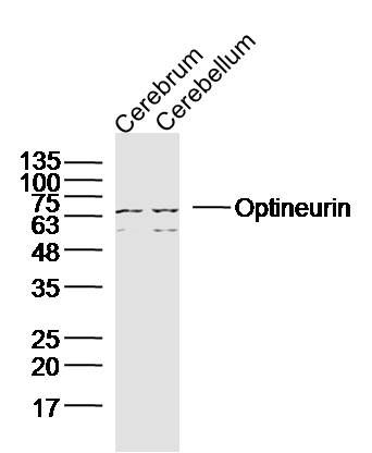 Sample:
Cerebrum (Mouse) Lysate at 40 ug Cerebellum (Mouse) Lysate at 40 ug Primary: Anti-Optineurin(bs-13658R)at 1/300 dilution Secondary: IRDye800CW Goat Anti-Rabbit IgG at 1/20000 dilution Predicted band size: 66kD Observed band size: 66kD  Paraformaldehyde-fixed, paraffin embedded (mouse testis); Antigen retrieval by boiling in sodium citrate buffer (pH6.0) for 15min; Block endogenous peroxidase by 3% hydrogen peroxide for 20 minutes; Blocking buffer (normal goat serum) at 37°C for 30min; Antibody incubation with (Optineurin) Polyclonal Antibody, Unconjugated (bs-13658R) at 1:400 overnight at 4°C, followed by a conjugated secondary (sp-0023) for 20 minutes and DAB staining.
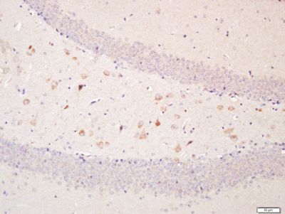 Paraformaldehyde-fixed, paraffin embedded (mouse brain); Antigen retrieval by boiling in sodium citrate buffer (pH6.0) for 15min; Block endogenous peroxidase by 3% hydrogen peroxide for 20 minutes; Blocking buffer (normal goat serum) at 37°C for 30min; Antibody incubation with (Optineurin) Polyclonal Antibody, Unconjugated (bs-13658R) at 1:400 overnight at 4°C, followed by a conjugated secondary (sp-0023) for 20 minutes and DAB staining.
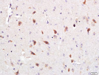 Paraformaldehyde-fixed, paraffin embedded (mouse brain); Antigen retrieval by boiling in sodium citrate buffer (pH6.0) for 15min; Block endogenous peroxidase by 3% hydrogen peroxide for 20 minutes; Blocking buffer (normal goat serum) at 37°C for 30min; Antibody incubation with (Optineurin) Polyclonal Antibody, Unconjugated (bs-13658R) at 1:400 overnight at 4°C, followed by a conjugated secondary (sp-0023) for 20 minutes and DAB staining.
|