Anti-Activated Notch1抗体
产品名称: Anti-Activated Notch1抗体
英文名称: Activated Notch1
产品编号: YB--20252R
产品价格: null
产品产地: 中国/美国
品牌商标: Ybscience
更新时间: 2023-08-17T10:29:50
使用范围: 科研使用
上海钰博生物科技有限公司
- 联系人 : 陈环环
- 地址 : 上海市沪闵路6088号龙之梦大厦8楼806室
- 邮编 : 200612
- 所在区域 : 上海
- 电话 : 183****2235
- 传真 : 021-60514606
- 邮箱 : shybio@126.com
Anti-Activated Notch1抗体
| 产品编号 | YB-20252R |
| 英文名称 | Activated Notch1 |
| 中文名称 | 活化的Notch1蛋白抗体 |
| 别 名 | Activated-notch1; Activated-notch 1; Activated notch 1; hN1; Neurogenic locus Notch homolog protein 1; activated Notch 1; activated Notch-1; Notch homolog 1 translocation associated (Drosophila); NOTCH1; TAN1; Translocation-associated Notch protein TAN-1; NOTC1_HUMAN. |
| 规格价格 | 100ul/1380元 购买 200ul/2200元 购买 大包装/询价 |
| 说 明 书 | 100ul 200ul |
| 研究领域 | 肿瘤 心血管 细胞生物 发育生物学 信号转导 干细胞 转录调节因子 表观遗传学 |
| 抗体来源 | Rabbit |
| 克隆类型 | Polyclonal |
| 交叉反应 | Human, Mouse, Rat, Dog, Pig, Cow, Rabbit, Guinea Pig, |
| 产品应用 | WB=1:500-2000 ELISA=1:500-1000 IHC-P=1:400-800 IHC-F=1:400-800 ICC=1:100-500 IF=1:100-500 (石蜡切片需做抗原修复) not yet tested in other applications. optimal dilutions/concentrations should be determined by the end user. |
| 分 子 量 | 89/271kDa |
| 细胞定位 | 细胞核 |
| 性 状 | Lyophilized or Liquid |
| 浓 度 | 1mg/ml |
| 免 疫 原 | KLH conjugated synthetic peptide derived from human N-terminal sequence of the cleaved Notch1 intracellular domain:1754-1800/2555 |
| 亚 型 | IgG |
| 纯化方法 | affinity purified by Protein A |
| 储 存 液 | 0.01M TBS(pH7.4) with 1% BSA, 0.03% Proclin300 and 50% Glycerol. |
| 保存条件 | Store at -20 °C for one year. Avoid repeated freeze/thaw cycles. The lyophilized antibody is stable at room temperature for at least one month and for greater than a year when kept at -20°C. When reconstituted in sterile pH 7.4 0.01M PBS or diluent of antibody the antibody is stable for at least two weeks at 2-4 °C. |
| PubMed | PubMed |
| 产品介绍 | background: This gene encodes a member of the Notch family. Members of this Type 1 transmembrane protein family share structural characteristics including an extracellular domain consisting of multiple epidermal growth factor-like (EGF) repeats, and an intracellular domain consisting of multiple, different domain types. Notch family members play a role in a variety of developmental processes by controlling cell fate decisions. The Notch signaling network is an evolutionarily conserved intercellular signaling pathway which regulates interactions between physically adjacent cells. In Drosophilia, notch interaction with its cell-bound ligands (delta, serrate) establishes an intercellular signaling pathway that plays a key role in development. Homologues of the notch-ligands have also been identified in human, but precise interactions between these ligands and the human notch homologues remain to be determined. This protein is cleaved in the trans-Golgi network, and presented on the cell surface as a heterodimer. This protein functions as a receptor for membrane bound ligands, and may play multiple roles during development. [provided by RefSeq, Jul 2008]. Function: Notch family members play a role in a variety of developmental processes by controlling cell fate decisions. The Notch signaling network is an evolutionarily conserved intercellular signaling pathway which regulates interactions between physically adjacent cells. The protein is cleaved in the trans-Golgi network, and presented on the cell surface as a heterodimer. This protein functions as a receptor for membrane bound ligands. Once the Notch extracellular domain interacts with a ligand, a protease called TACE (Tumor Necrosis Factor Alpha Converting Enzyme) cleaves the Notch protein just outside the membrane. This releases the extracellular portion of Notch, which continues to interact with the ligand. The ligand plus the Notch extracellular domain is then endocytosed by the ligand expressing cell. After this first cleavage, an enzyme called gamma-secretase cleaves the remaining part of the Notch protein just inside the inner leaflet of the cell membrane. This releases the intracellular portion of the Notch protein, which then moves to the nucleus and causes various genes to be expressed. There are many other proteins involved in the intracellular portion of the Notch signalling cascade. Subunit: Heterodimer of a C-terminal fragment N(TM) and an N-terminal fragment N(EC) which are probably linked by disulfide bonds. Interacts with DNER, DTX1, DTX2 and RBPJ/RBPSUH. Also interacts with MAML1, MAML2 and MAML3 which act as transcriptional coactivators for NOTCH1. The activated membrane-bound form interacts with AAK1 which promotes NOTCH1 stabilization. Forms a trimeric complex with FBXW7 and SGK1. Interacts with HIF1AN. HIF1AN negatively regulates the function of notch intracellular domain (NICD), accelerating myogenic differentiation. Subcellular Location: Cell membrane; Single-pass type I membrane protein. Notch 1 intracellular domain: Nucleus. Note=Following proteolytical processing NICD is translocated to the nucleus. Tissue Specificity: In fetal tissues most abundant in spleen, brain stem and lung. Also present in most adult tissues where it is found mainly in lymphoid tissues. Post-translational modifications: Synthesized in the endoplasmic reticulum as an inactive form which is proteolytically cleaved by a furin-like convertase in the trans-Golgi network before it reaches the plasma membrane to yield an active, ligand-accessible form. Cleavage results in a C-terminal fragment N(TM) and a N-terminal fragment N(EC). Following ligand binding, it is cleaved by TNF-alpha converting enzyme (TACE) to yield a membrane-associated intermediate fragment called notch extracellular truncation (NEXT). Following endocytosis, this fragment is then cleaved by presenilin dependent gamma-secretase to release a notch-derived peptide containing the intracellular domain (NICD) from the membrane (By similarity). Phosphorylated (By similarity). O-glycosylated on the EGF-like domains. Contains both O-linked fucose and O-linked glucose. Ubiquitinated; undergoes 'Lys-29'-linked polyubiquitination catalyzed by ITCH. Monoubiquitination at Lys-1759 is required for activation by gamma-secretase cleavage, it promotes interaction with AAK1, which stabilizes it. Deubiquitination by EIF3F is necessary for nuclear import of activated Notch. Hydroxylated at Asn-1955 by HIF1AN. Hydroxylated at Asn-2022 by HIF1AN (By similarity). Hydroxylation reduces affinity for HI1AN and may thus indirectly modulate negative regulation of NICD. DISEASE: Defects in NOTCH1 are a cause of aortic valve disease 1 (AOVD1) [MIM:109730]. A common defect in the aortic valve in which two rather than three leaflets are present. It is often associated with aortic valve calcification and insufficiency. In extreme cases, the blood flow may be so restricted that the left ventricle fails to grow, resulting in hypoplastic left heart syndrome. Similarity: Belongs to the NOTCH family. Contains 5 ANK repeats. Contains 36 EGF-like domains. Contains 3 LNR (Lin/Notch) repeats. SWISS: P46531 Gene ID: 4851 Database links: Entrez Gene: 4851 Human Entrez Gene: 18128 Mouse Entrez Gene: 767866 Cow Omim: 190198 Human SwissProt: P46531 Human SwissProt: Q01705 Mouse Unigene: 495473 Human nigene: 290610 Mouse Important Note: This product as supplied is intended for research use only, not for use in human, therapeutic or diagnostic applications. |
| 产品图片 |
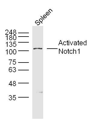 Sample:spleen (mouse) Lysate at 40 ug
Primary: Anti-Activated Notch1(bs-20252R)at 1/300 dilution Secondary: IRDye800CW Goat Anti-RabbitIgG at 1/20000 dilution Predicted band size: 86kD Observed band size: 107kD 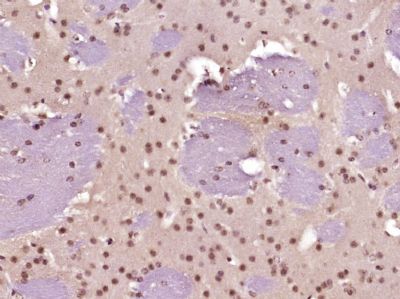 Paraformaldehyde-fixed, paraffin embedded (Mouse brain); Antigen retrieval by boiling in sodium citrate buffer (pH6.0) for 15min; Block endogenous peroxidase by 3% hydrogen peroxide for 20 minutes; Blocking buffer (normal goat serum) at 37°C for 30min; Antibody incubation with (Activated Notch1) Polyclonal Antibody, Unconjugated (bs-20252R) at 1:400 overnight at 4°C, followed by operating according to SP Kit(Rabbit) (sp-0023) instructions and DAB staining.
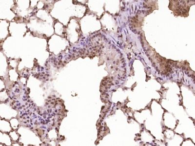 Paraformaldehyde-fixed, paraffin embedded (Mouse lung); Antigen retrieval by boiling in sodium citrate buffer (pH6.0) for 15min; Block endogenous peroxidase by 3% hydrogen peroxide for 20 minutes; Blocking buffer (normal goat serum) at 37°C for 30min; Antibody incubation with (Activated Notch1) Polyclonal Antibody, Unconjugated (bs-20252R) at 1:400 overnight at 4°C, followed by operating according to SP Kit(Rabbit) (sp-0023) instructions and DAB staining.
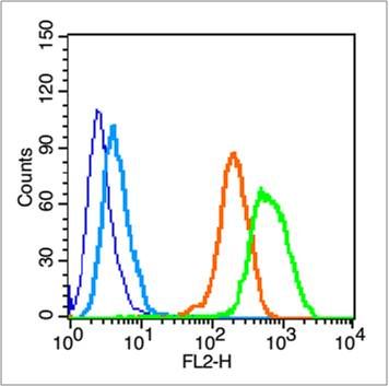 Blank control (blue line): Hela (fixed with 70% ethanol overnight at 4℃ and then permeabilized with 90% ice-cold methanol for 30 min on ice).
Primary Antibody (green line): Rabbit Anti-Activated Notch1 antibody (bs-20252R),Dilution: 1μg /10^6 cells; Isotype Control Antibody (orange line): Rabbit IgG . Secondary Antibody (white blue line): Goat anti-rabbit IgG-PE, Dilution: 1μg /test. |