Anti-ALK抗体
产品名称: Anti-ALK抗体
英文名称: ALK
产品编号: YB--0097R
产品价格: null
产品产地: 中国/美国
品牌商标: Ybscience
更新时间: 2023-08-17T10:29:50
使用范围: 科研使用
上海钰博生物科技有限公司
- 联系人 : 陈环环
- 地址 : 上海市沪闵路6088号龙之梦大厦8楼806室
- 邮编 : 200612
- 所在区域 : 上海
- 电话 : 183****2235
- 传真 : 021-60514606
- 邮箱 : shybio@126.com
Anti-ALK抗体
| 产品编号 | YB-0097R |
| 英文名称 | ALK |
| 中文名称 | 间变型淋巴瘤激酶抗体 |
| 别 名 | ALK tyrosine kinase receptor; ALK tyrosine kinase receptor precursor; ALK/EML4 fusion gene, included; ALK/NPM1 fusion gene, included; anaplastic lymphoma kinase (Ki-1); Anaplastic lymphoma kinase; Anaplastic lymphoma kinase Ki 1; Anaplastic lymphoma kinase Ki1; Anaplastic lymphoma kinase p80; CD 246; CD246; CD-246; CD246 antigen; EC 2.7.10.1; Ki 1; Ki1; NBLST3; Tcrz; TFG/ALK. |
| 规格价格 | 100ul/1380元 购买 200ul/2200元 购买 大包装/询价 |
| 说 明 书 | 100ul 200ul |
| 研究领域 | 免疫学 神经生物学 信号转导 生长因子和激素 转录调节因子 激酶和磷酸酶 细胞类型标志物 b-淋巴细胞 肿瘤细胞生物标志物 |
| 抗体来源 | Rabbit |
| 克隆类型 | Polyclonal |
| 交叉反应 | Human, Mouse, Rat, Dog, Cow, Horse, |
| 产品应用 | ELISA=1:500-1000 IHC-P=1:400-800 IHC-F=1:400-800 Flow-Cyt=1μg /test IF=1:100-500 (石蜡切片需做抗原修复) not yet tested in other applications. optimal dilutions/concentrations should be determined by the end user. |
| 分 子 量 | 174kDa |
| 细胞定位 | 细胞膜 |
| 性 状 | Lyophilized or Liquid |
| 浓 度 | 1mg/ml |
| 免 疫 原 | KLH conjugated synthetic peptide derived from human CD246:329-342/1620 |
| 亚 型 | IgG |
| 纯化方法 | affinity purified by Protein A |
| 储 存 液 | 0.01M TBS(pH7.4) with 1% BSA, 0.03% Proclin300 and 50% Glycerol. |
| 保存条件 | Store at -20 °C for one year. Avoid repeated freeze/thaw cycles. The lyophilized antibody is stable at room temperature for at least one month and for greater than a year when kept at -20°C. When reconstituted in sterile pH 7.4 0.01M PBS or diluent of antibody the antibody is stable for at least two weeks at 2-4 °C. |
| PubMed | PubMed |
| 产品介绍 | background: This gene encodes a receptor tyrosine kinase, which belongs to the insulin receptor superfamily. This protein comprises an extracellular domain, an hydrophobic stretch corresponding to a single pass transmembrane region, and an intracellular kinase domain. It plays an important role in the development of the brain and exerts its effects on specific neurons in the nervous system. This gene has been found to be rearranged, mutated, or amplified in a series of tumours including anaplastic large cell lymphomas, neuroblastoma, and non-small cell lung cancer. The chromosomal rearrangements are the most common genetic alterations in this gene, which result in creation of multiple fusion genes in tumourigenesis, including ALK (chromosome 2)/EML4 (chromosome 2), ALK/RANBP2 (chromosome 2), ALK/ATIC (chromosome 2), ALK/TFG (chromosome 3), ALK/NPM1 (chromosome 5), ALK/SQSTM1 (chromosome 5), LK/KIF5B (chromosome 10), ALK/CLTC (chromosome 17), ALK/TPM4 (chromosome 19), and ALK/MSN (chromosome X).[provided by RefSeq, Jan 2011]. Function: Neuronal orphan receptor tyrosine kinase that is essentially and transiently expressed in specific regions of the central and peripheral nervous systems and plays an important role in the genesis and differentiation of the nervous system. Transduces signals from ligands at the cell surface, through specific activation of the mitogen-activated protein kinase (MAPK) pathway. Phosphorylates almost exclusively at the first tyrosine of the Y-x-x-x-Y-Y motif. Following activation by ligand, ALK induces tyrosine phosphorylation of CBL, FRS2, IRS1 and SHC1, as well as of the MAP kinases MAPK1/ERK2 and MAPK3/ERK1. Acts as a receptor for ligands pleiotrophin (PTN), a secreted growth factor, and midkine (MDK), a PTN-related factor, thus participating in PTN and MDK signal transduction. Subunit: Homodimer. Homodimerizes when bound to ligand. Interacts with FRS2, IRS1, MDK, PTN and SHC1. Interacts with CBL, PIK3R1 and PLCG1. Subcellular Location: Cell membrane; Single-pass type I membrane protein. Note=Membrane attachment was crucial for promotion of neuron-like differentiation and cell proliferation arrest through specific activation of the MAP kinase pathway. Tissue Specificity: Expressed in brain and CNS. Also expressed in the small intestine and testis, but not in normal lymphoid cells. Post-translational modifications: Phosphorylated at tyrosine residues by autocatalysis, which activates kinase activity. In cells not stimulated by a ligand, receptor protein tyrosine phosphatase beta and zeta complex (PTPRB/PTPRZ1) dephosphorylates ALK at the sites in ALK that are undergoing autophosphorylation through autoactivation. N-glycosylated. DISEASE: Note=A chromosomal aberration involving ALK is found in a form of non-Hodgkin lymphoma. Translocation t(2;5)(p23;q35) with NPM1. The resulting chimeric NPM1-ALK protein homodimerize and the kinase becomes constitutively activated. The constitutively active fusion proteins are responsible for 5-10% of non-Hodgkin Note=A chromosomal aberration involving ALK is associated with inflammatory myofibroblastic tumors (IMTs). Translocation t(2;11)(p23;p15) with CARS; translocation t(2;4)(p23;q21) with SEC31A. Similarity: Belongs to the protein kinase superfamily. Tyr protein kinase family. Insulin receptor subfamily. Contains 1 LDL-receptor class A domain. Contains 2 MAM domains. Contains 1 protein kinase domain. SWISS: Q9UM73 Gene ID: 238 Database links:
Entrez Gene: 238 Human Entrez Gene: 11682 Mouse Entrez Gene: 266802 Rat Omim: 105590 Human SwissProt: Q9UM73 Human SwissProt: P97793 Mouse Unigene: 654469 Human Unigene: 311854 Mouse Unigene: 201918 Rat
Important Note: This product as supplied is intended for research use only, not for use in human, therapeutic or diagnostic applications. ALK蛋白在细胞生长调控中起重要作用,主要表达在神经细胞,白细胞癌基因蛋白。 ALK p80是多向性生长因子酪氨酸激酶受体蛋白, 间变性大细胞淋巴瘤伴有t(2;5) (p23;q35)染色体的易位,易位后的细胞基因表达分子量为80kD,常伴有CD30阳性,ALK p80阳性的间变性大细胞淋巴瘤预后好于阴性病例。 主要用于间变性大细胞淋巴瘤与何杰金氏淋巴瘤的鉴别诊断,间变性大细胞淋巴瘤中阳性率大约为70%左右,还可以用于骨髓中间变性大细胞淋巴瘤的诊断. |
| 产品图片 |
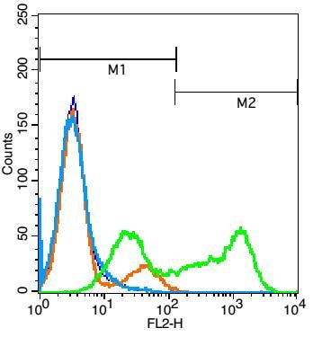 Blank control: Jurkat cells(blue).
Primary Antibody:Rabbit Anti-ALK antibody(bs-0097R), Dilution: 1μg in 100 μL 1X PBS containing 0.5% BSA; Isotype Control Antibody: Rabbit IgG(orange) ,used under the same conditions ); Secondary Antibody: Goat anti-rabbit IgG-PE(white blue), Dilution: 1:200 in 1 X PBS containing 0.5% BSA. Protocol The cells were fixed with 2% paraformaldehyde (10 min) . Primary antibody (bs-0097R, 1μg /1x10^6 cells) were incubated for 30 min on the ice, followed by 1 X PBS containing 0.5% BSA + 1 0% goat serum (15 min) to block non-specific protein-protein interactions. Then the Goat Anti-rabbit IgG/PE antibody was added into the blocking buffer mentioned above to react with the primary antibody at 1/200 dilution for 30 min on ice. Acquisition of 20,000 events was performed. |