Anti-TGFBI/FITC Conjugated抗体
产品名称: Anti-TGFBI/FITC Conjugated抗体
英文名称: Anti-TGFBI/FITC
产品编号: YB--7443R-FITC
产品价格: null
产品产地: 中国/美国
品牌商标: Ybscience
更新时间: 2023-08-17T10:29:50
使用范围: 科研使用
上海钰博生物科技有限公司
- 联系人 : 陈环环
- 地址 : 上海市沪闵路6088号龙之梦大厦8楼806室
- 邮编 : 200612
- 所在区域 : 上海
- 电话 : 183****2235
- 传真 : 021-60514606
- 邮箱 : shybio@126.com
Anti-TGFBI/FITC Conjugated抗体
| 产品编号 | YB-7443R-FITC |
| 英文名称 | Anti-TGFBI/FITC |
| 中文名称 | FITC标记的角膜上皮蛋白TGFBI抗体 |
| 别 名 | AI181842; AI747162; Beta ig; Beta ig h3; Beta ig-h3; BGH3_HUMAN; Big h3; BIGH3; CDB1; CDG2; CDGG1; CSD; CSD1; CSD2; CSD3; EBMD; Kerato epithelin; Kerato-epithelin; LCD1. |
| 规格价格 | 100ul/2980元 购买 大包装/询价 |
| 说 明 书 | 100ul |
| 研究领域 | 细胞生物 发育生物学 神经生物学 信号转导 干细胞 生长因子和激素 |
| 抗体来源 | Rabbit |
| 克隆类型 | Polyclonal |
| 交叉反应 | Human, Mouse, Rat, Dog, Pig, Cow, Horse, Rabbit, Sheep, |
| 产品应用 | ICC=1:50-200 IF=1:50-200 not yet tested in other applications. optimal dilutions/concentrations should be determined by the end user. |
| 分 子 量 | 72kDa |
| 性 状 | Lyophilized or Liquid |
| 浓 度 | 2mg/1ml |
| 免 疫 原 | KLH conjugated synthetic peptide derived from human TGFBI |
| 亚 型 | IgG |
| 纯化方法 | affinity purified by Protein A |
| 储 存 液 | 0.01M TBS(pH7.4) with 1% BSA, 0.03% Proclin300 and 50% Glycerol. |
| 保存条件 | Store at -20 °C for one year. Avoid repeated freeze/thaw cycles. The lyophilized antibody is stable at room temperature for at least one month and for greater than a year when kept at -20°C. When reconstituted in sterile pH 7.4 0.01M PBS or diluent of antibody the antibody is stable for at least two weeks at 2-4 °C. |
| 产品介绍 | background: This gene encodes an RGD-containing protein that binds to type I, II and IV collagens. The RGD motif is found in many extracellular matrix proteins modulating cell adhesion and serves as a ligand recognition sequence for several integrins. This protein plays a role in cell-collagen interactions and may be involved in endochondrial bone formation in cartilage. The protein is induced by transforming growth factor-beta and acts to inhibit cell adhesion. Mutations in this gene are associated with multiple types of corneal dystrophy. [provided by RefSeq, Jul 2008] Function: Binds to type I, II, and IV collagens. This adhesion protein may play an important role in cell-collagen interactions. In cartilage, may be involved in endochondral bone formation. Subcellular Location: Secreted > extracellular space > extracellular matrix. May be associated both with microfibrils and with the cell surface. Tissue Specificity: Highly expressed in the corneal epithelium. Post-translational modifications: Gamma-carboxyglutamate residues are formed by vitamin K dependent carboxylation. These residues are essential for the binding of calcium. DISEASE: Defects in TGFBI are the cause of epithelial basement membrane corneal dystrophy (EBMD) [MIM:121820]; also known as Cogan corneal dystrophy or map-dot-fingerprint type corneal dystrophy. EBMD is a bilateral anterior corneal dystrophy characterized by grayish epithelial fingerprint lines, geographic map-like lines, and dots (or microcysts) on slit-lamp examination. Pathologic studies show abnormal, redundant basement membrane and intraepithelial lacunae filled with cellular debris. Although this disorder usually is not considered to be inherited, families with autosomal dominant inheritance have been identified. Defects in TGFBI are the cause of corneal dystrophy Groenouw type 1 (CDGG1) [MIM:121900]; also known as corneal dystrophy granular type. Inheritance is autosomal dominant. Corneal dystrophies show progressive opacification of the cornea leading to severe visual handicap. Defects in TGFBI are the cause of corneal dystrophy lattice type 1 (CDL1) [MIM:122200]. Inheritance is autosomal dominant. Defects in TGFBI are a cause of corneal dystrophy Thiel-Behnke type (CDTB) [MIM:602082]; also known as corneal dystrophy of Bowman layer type 2 (CDB2). Defects in TGFBI are the cause of Reis-Buecklers corneal dystrophy (CDRB) [MIM:608470]; also known as corneal dystrophy of Bowman layer type 1 (CDB1). Defects in TGFBI are the cause of lattice corneal dystrophy type 3A (CDL3A) [MIM:608471]. CDL3A clinically resembles to lattice corneal dystrophy type 3, but differs in that its age of onset is 70 to 90 years. It has an autosomal dominant inheritance pattern. Defects in TGFBI are the cause of Avellino corneal dystrophy (ACD) [MIM:607541]. ACD could be considered a variant of granular dystrophy with a significant amyloidogenic tendency. Inheritance is autosomal dominant. Similarity: Contains 1 EMI domain. Contains 4 FAS1 domains. Database links: Entrez Gene: 7045 Human Entrez Gene: 21810 Mouse Entrez Gene: 116487 Rat Omim: 601692 Human SwissProt: Q15582 Human SwissProt: P82198 Mouse Unigene: 369397 Human Unigene: 14455 Mouse Important Note: This product as supplied is intended for research use only, not for use in human, therapeutic or diagnostic applications. |
| 产品图片 |
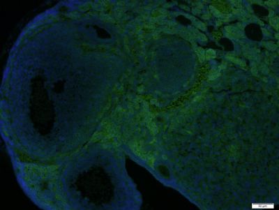 Paraformaldehyde-fixed, paraffin embedded (Mouse ovary); Antigen retrieval by boiling in sodium citrate buffer (pH6.0) for 15min; Block endogenous peroxidase by 3% hydrogen peroxide for 20 minutes; Blocking buffer (normal goat serum) at 37°C for 30min; Incubation: Anti-TGFBI Antibody, conjugated (bs-7443R-FITC) 1:400, 1.5 hours at 37°C; DAPI (5ug/ml, blue, C-0033) was used to stain the cell nuclei.
 Paraformaldehyde-fixed, paraffin embedded (Rat colon); Antigen retrieval by boiling in sodium citrate buffer (pH6.0) for 15min; Block endogenous peroxidase by 3% hydrogen peroxide for 20 minutes; Blocking buffer (normal goat serum) at 37°C for 30min; Incubation: Anti-TGFBI Antibody, conjugated (bs-7443R-FITC) 1:400, 1.5 hours at 37°C; DAPI (5ug/ml, blue, C-0033) was used to stain the cell nuclei.
|