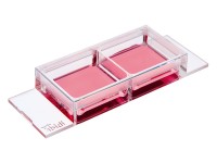
μ培养载玻片 2孔
产品名称: μ培养载玻片 2孔
英文名称: ibidi
产品编号: 80296 80292 80293 80294 80295 80291
产品价格: 0
产品产地: 德国
品牌商标: ibidi
更新时间: 2023-08-17T17:25:28
使用范围: null
- 联系人 : 黄燕飞
- 地址 : 上海市松江区曹农路5弄40号
- 邮编 : 201100
- 所在区域 : 上海
- 电话 : 177****7476
- 传真 : 021-57790918
- 邮箱 : 3680024208@qq.com
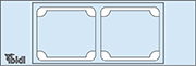
μ-Slide 2 well Ph+/80296 80292 80293 80294 80295 80291
|
货号 |
产品名称 |
规格(个/盒) |
|
80296 |
μ-Slide 2孔Ph+腔室载玻片,ibiTreat底部处理 |
15 |
|
80292 |
μ-Slide 2孔Ph+腔室载玻片,Collagen IV底部处理 |
15 |
|
80293 |
μ-Slide 2孔Ph+腔室载玻片,Fibronectin底部处理 |
15 |
|
80294 |
μ-Slide 2孔Ph+腔室载玻片,Poly-L-Lysine底部处理 |
15 |
|
80295 |
μ-Slide 2孔Ph+腔室载玻片,Poly-D-Lysine底部处理 |
15 |
|
80291 |
μ-Slide 2孔Ph+腔室载玻片,无包被 |
15 |
|
80297 |
μ-Slide 2孔Ph+腔室载玻片,玻璃底 |
15 |
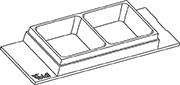
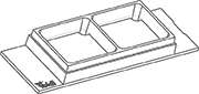
μ培养载玻片 2孔Ph+
一个开放的两孔培养载玻片,并带有一个特殊中间板,非常适用于相差和的荧光显微镜
整个培养孔具有的相差-没有弯月面现象
体化小室,既可用于细胞培养,也可用于显微镜成像
使用高分辨率显微镜可以通过玻璃片底部进行样品观察
没有盖玻片,不会泄露,用于快速,简单的免疫荧光。
应用
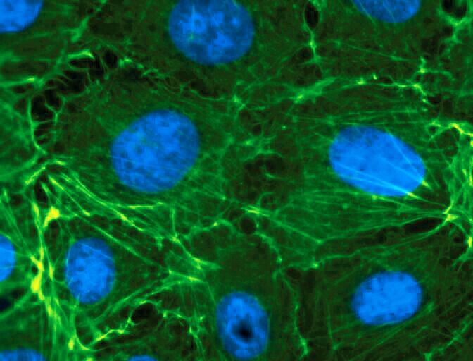
细胞培养以及细胞培养物的显微镜观察。
转染分析
活细胞和固定细胞的免疫荧光显微镜观察
长时间段下活细胞成像
规格
|
孔数量 |
2 |
带盖子高度 |
10.8 mm |
|
孔尺寸大小 |
2.2 x 23.3 x 9.3 |
每孔生成面积 |
4.8 cm2 |
|
每孔体积 |
1500 μl |
每孔包被面积 |
11.4 cm2 |
|
底部 ibidi 标准底部 |
|||
技术特点
开放式载玻片,具有两个独立的培养孔
每个培养孔上的中间板可以避免弯液面形成
从两侧缝隙很容易加液,无气泡形成,
适用于显微镜的光学成像特质
兼容染色,固定
采用佳细胞粘附的表面处理Ibitreat
生物兼容性塑料生产,无胶水,不泄露
相差显微镜是常用的,细胞培养中的透射光学技术。使用相差显微镜时,把两个相位环彼此调节好是非常重要的。在开放式培养孔中,空气-水交界面处的弯月牙就像一个棱镜折射出光路。这个会误校相位环,造成显微镜图像的对比性较差。
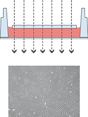
使用μ- 2孔ph+培养载玻片减少了弯月牙,无论孔在哪个位置,整个光学系统都是平行的
μ-Slide 2 well μ-Slide 2 well Ph+

The μ-Slide 2 well Ph+ (Phase Contrast +) is designed for excellent optical quality, especially for cell culture when normal phase contrast microscopy is being used. Opposed to the classic μ-Slide 2 well, the Ph+ version provides a special intermediate plate in the center of the well. This plate flattens the meniscus that disturbs the phase contrast effect in normal open wells. Openings near the corners provide access to the wells for easy filling and aspiration of liquids.
This innovative technique supports meniscus-free phase contrast microscopy in a very convenient manner.
Cross Section Through a Well of the μ-Slide 2 well Ph+ with a Transmitted Light Beam Path
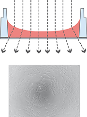
The illustration on the left shows the perturbing effect of a meniscus. Light is refracted on the air-water-interface, leading to poor contrast in microscopy. Only the small center part exhibits satisfying phase contrast.
Working with the μ-Slide 2 well Ph+ diminishes the meniscus and increases the area of nicely contrasted cells. This nice contrast is due to the parallel beam path that is created by the plate.