德国ibidi μ-Slide ibiPore Transwell细胞侵袭-85036
产品名称: 德国ibidi μ-Slide ibiPore Transwell细胞侵袭-85036
英文名称: 德国ibidi
产品编号: 85036
产品价格: 面议
产品产地: 上海
品牌商标: 德国ibidi
更新时间: 2024-04-03T11:11:46
使用范围: null
- 联系人 : 黄燕飞
- 地址 : 上海市松江区曹农路5弄40号
- 邮编 : 201100
- 所在区域 : 上海
- 电话 : 177****7476
- 传真 : 021-57790918
- 邮箱 : 3680024208@qq.com
货号
产品名称
规格(个/盒)
85016
μ-Slide ibiPore培养载玻片,可视化Transwell 0.5μm/20%, ibiTreat底部处理
10
85026
μ-Slide ibiPore培养载玻片,可视化Transwell 3.0μm/5%, ibiTreat底部处理
10
85036
μ-Slide ibiPore培养载玻片,可视化Transwell 5.0μm/5%, ibiTreat底部处理
10
85046
μ-Slide ibiPore培养载玻片,可视化Transwell 8.0μm/5%, ibiTreat底部处理
10
850?6-S
以上μ-Slide ibiPore培养载玻片,小包装, ibiTreat底部处理
2
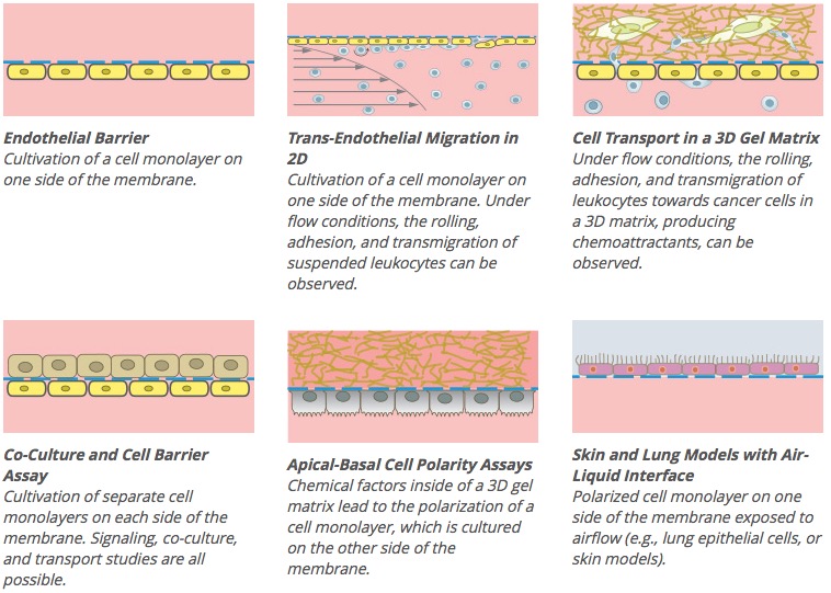
应用:
1.流动状态下跨内皮细胞迁移
2.2D或者3D凝胶内细胞层共培养和运输
3.顶部-基底细胞急性分析
4.液体-空气接触的皮肤和肺部模型
5.顶部-基底梯度的细胞屏障模型
6.基于过滤系统和孔状膜的细胞迁移分析
技术特点:
1.SiMPore’s G-FLAT™ 微孔径玻璃膜
2.中间具有玻璃光学膜的跨通道结构
3.与盖玻片相比,具有较好的光学特质
4.孔径大小0.5μm,3μm,5μm,8μm
5.膜厚度0.3μm(300nm)
6.物镜工作距离>0.5mm
7.兼容ibidi泵系统
8.特定剪切力大小和剪切力速率水平
(详情点击: Application Note 11)
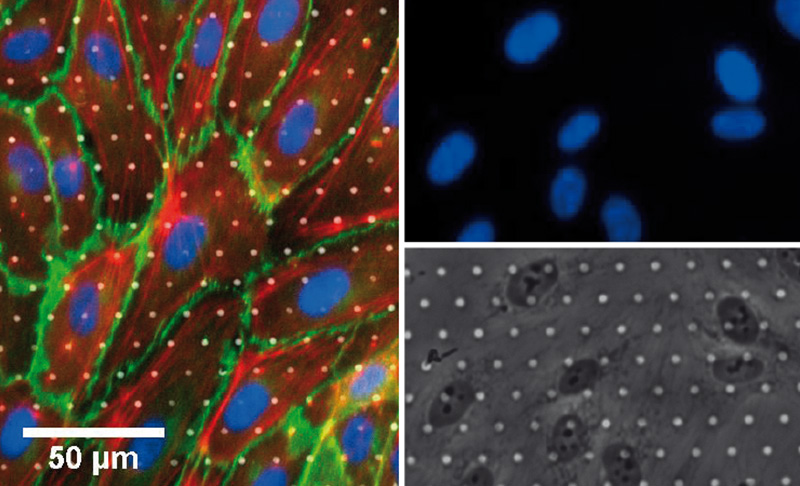
Endothelial cells on 3 μm ibiPore membrane. Immunofluorescence overlay image of phase contrast, DAPI (blue), VE-Cadherins (green), F-Actin (red).
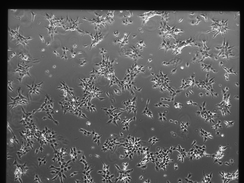
Fibroblast cells on 3 μm ibiPore membrane.
Phase contrast image, objective lens 4x.
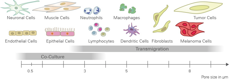
应用建议: 孔径 & 孔密度
| 膜类型 | 应用 | 细胞种类案例 |
| 0.5 μm pores, high porosity (20%) | Permeability and transport studies, co-culture models. With this model, cell migration is not possible. | Lung cells, epithelial cells |
| 3.0 μm pores, low porosity (5%) | Transendothelial migration | Leukocytes (neutrophils, T-cells) |
| 5.0 μm pores, low porosity (5%) | Invasion, migration | Monocytes, macrophages, lymphocytes |
| 8.0 μm pores, low porosity (5%) | Invasion, migration | Tumor and cancer cells, endothelial and epithelial cells, fibroblasts, osteoblasts, melanoma, glioma |
越大的细胞 – 建议使用越大的孔径。
不同应用的建议孔径:
μ-Slide ibiPore
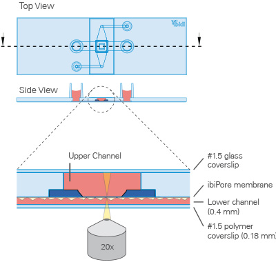
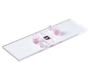
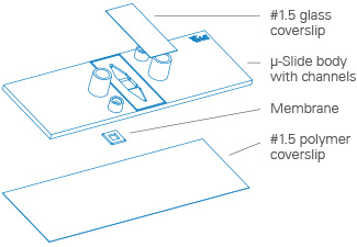
μ-Slide ibiPore 细胞侵袭载玻片结构
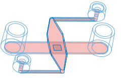
交叉通道结构和中间ibiPore膜结构的3D示意图
应用实例
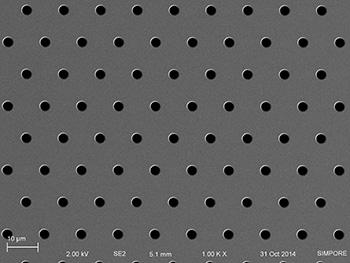
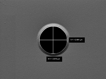
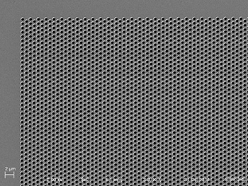
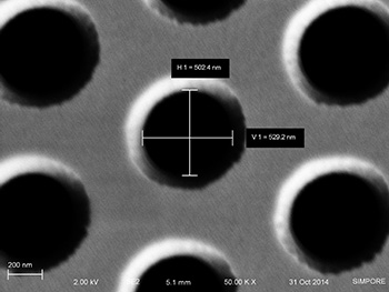
扫描电镜下的多孔玻璃膜
3微米孔
0.5微米孔