软骨灌流离体剪切应力组织工程3D培养生物反应器
产品名称: 软骨灌流离体剪切应力组织工程3D培养生物反应器
英文名称:
产品编号: TEB
产品价格: 0
产品产地: 西班牙
品牌商标: ebers
更新时间: null
使用范围: null
- 联系人 :
- 地址 : 中国上海上海上海市
- 邮编 : 201112
- 所在区域 : 上海
- 电话 : 180****9478 点击查看
- 传真 : 点击查看
- 邮箱 : brightzhou@micoforce.com
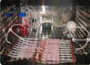
灌流软骨生物反应器(perfusion cartilage bioreactors)原理为连续添加培养液至培养舱,同时也吸出相同体积的原有培养液,使槽内培养液体积维持一定,养分也能维持在细胞生长所需范围以上,细胞代谢产物则不会堆积太多。培养时将已完成种植细胞的支架固定于灌流系统中,培养基直接流过支架之孔隙,流体可在支架的截面均匀分布,让接种的细胞随之均匀注入多孔支架中贴附培养。灌流可是密闭系统,经由另一个培养液容器再循环回到原先之培养槽。此外,灌流反应器克服了机械混合产生的剪应力,且不会产生中间坏死的情况。若对细胞的周围环境包括pH值、温度、培养液的营养成分、代谢产物等,作精确的监测和控制,培养细胞的密度及质量可以提高。 EBER Effect of the Shear Stress in the Human Bone Marrow Mesenchymal Stem Cell Behavior Clara Alcaine1,2, Sonia Santander1,2, Ignacio Ochoa1,2, Jose Manuel Garcia-Aznar1,2, Manuel Doblare1,2 Corresponding Author: iochgar@unizar.es 1 Group of Structural Mechanics and Materials Modelling (GEMM). Aragón Institute of Engineering Research (I3A). Universidad de Zaragoza, Spain 2 Centro de Investigación Biomédica en Red en Bioingeniería, Biomateriales y Nanomedicina (CIBER-BBN) Aragón Institute of Health Sciences, Spain Introduction Shear stress has been previously reported to be a powerful differentiation stimuli (1,2). However, other effects (proliferation, migration) on Human Bone Marrow Mesenchymal Stem Cells (MSCs) have not been deeply studied. Our main goal is to determine if the shear stress is able to affect not only differentiation but also other important cell processes such as cell proliferation, migration or cell adhesion. Materials and Methods Bone Marrow Mesenchymal Stem Cells have been seeded in μ-Slide I flow kit (Ibidi) and cultured under a shear stress of 5 dynes /cm2 in a perfusion bioreactor (TEB-1000, EBERS) for 7 days. Proliferation was determined after cell counting and apoptosis (Annexin V) was measured with flow citometry techniques. Migration experiments were performed in a multidimensional microscope for 24 hours. Inmunofluorescent staining to determine the cell area and cytoskeleton organization was carried out in a confocal microscope. Expression of the cell adhesion molecules was determined by RTPCR. ANOVA and t-student tests were carried out to determine statistical differences. Results Significant differences in cell proliferation have been observed after 7 days between static and shear stimulated cells (Fig.1). However, no significant differences in apoptosis were obtained between both studied groups (Fig.2). Differences in cell area, speed of migration, cytoskeleton organization and adhesion molecules has been also observed. In a future, we will try to determine the effect of different shear stress ratios on the previously described variables.