经典文献回顾-ex Foxp3 Th17细胞的哲学独白
“我是谁?我从哪里来?我要到哪里去” 是柏拉图提出的经典哲学三大问,小编每天早上起床前都要用这个问题叫醒我沉睡的灵魂。在本期带来的经典文献回顾中,请跟随本文作者一起听一听ex Foxp3 TH17细胞的深刻哲学独白吧!
一、故事背景
自身免疫性疾病通常由调节性T细胞(Treg细胞)和产生干扰素17的辅助T细胞( TH17细胞)失衡所导致,严重影响着人类的健康。但TH17细胞的来源仍然有很多未知,于是,主人公ex Foxp3 TH17细胞的独白在自身免疫性关节炎中开始了。
二、哲学独白
第一问:我从哪里来?
ex Foxp3 TH17细胞闷闷不乐托腮思考:我作为TH17细胞的一员,感觉跟其他TH17细胞小伙伴儿们不是特别合群,常常有不一样的感觉,我是从哪里来的呢?
让我从头开始捋一捋,先找找影响自身免疫性关节炎的关键人物:表达Foxp3的Treg细胞已经知道是抑制免疫应答的关键角色,小鼠缺失Foxp3会产生自身免疫疾病,持续表达则会抑制自身免疫,可见Foxp3表达的稳定性是两者平衡的重要因素。
那么Foxp3的稳定性对自身免疫关节炎有什么影响呢? Treg细胞的发展和功能受IL-2调控,Foxp3+T细胞由Foxp3+-稳定CD25(IL-2Rα)hi和Foxp3+-不稳定CD25 lo细胞群组成。作者将CD25hiFoxp3+CD4+细胞和CD25loFoxp3+CD4+细胞(来源于未处理的DBA/1 Foxp3hCD2小鼠,hCD2作为标签指示Foxp3表达情况)转移到胶原诱导型关节炎(collagen-induced arthritis ,CIA)疾病模型小鼠上,测定临床评分及踝关节损伤情况,发现CD25hiFoxp3+CD4+细胞与对照组及CD25loFoxp3+CD4+细胞相比减轻了关节肿胀和损伤等症状(图1a,b)。将细胞经CFSE染料标记后再转移,结果显示在脾脏和引流淋巴结(dLNs)中CD25loFoxp3+CD4+细胞丢失了部分Foxp3的表达(30-50%)(图1c)。由此可见, CD25loFoxp3+CD4+细胞因为丢失了Foxp3而没有办法缓解发炎和骨损伤症状。思考到这里,我忍不住想问了:丢掉Foxp3的CD25loFoxp3+CD4+细胞去哪了?
别着急,我们继续追踪细胞更长时间的发展, 将total Foxp3 + 、CD25hiFoxp3+CD4+和CD25loFoxp3+CD4+细胞(来源于B6.Ly5.1 Foxp3hCD2小鼠,donor)转到C57BL/6 Ly5.2 关节炎小鼠(host)模型中,检测Foxp3和IL17表达, CD25loFoxp3+CD4+细胞失去部分Foxp3表达,产生了更高的IL-17 (图1d,e)。且检测到CD25loFoxp3+ 来源的Ly5.1+Foxp3-CD4+ T细胞比naïve CD4+ T细胞来源的有更多IL-17和CCR6的表达(两者为TH17细胞marker)。这下清楚了,原来这些CD25loFoxp3+CD4+细胞丢失了Foxp3以后就变成我( ex Foxp3 TH17细胞)了,我是从CD25loFoxp3+CD4+细胞分化而来呀。

Figure 1 CD25loFoxp3+ T cells are unstable Foxp3+ T cells that convert to TH17 cells under arthritic conditions. (a,b) Clinical score (a) and microcomputed tomography analysis of calcaneus in the ankle joints (b) of immunized DBA/1 mice adoptively transferred with 5 × 10⁵ CD25hi (a, n = 3; b, n = 6) or CD25lo (a, n = 5; b, n = 10) Foxp3+CD4+ T cells purified from untreated DBA/1 Foxp3hCD2 mice. (c) Frequency of Foxp3+ cells in CFSE+ donor-derived T cells. Representative data of five mice are shown. (d,e) Results from the immunized C57BL/6 Ly5.2 mice that were adoptively transferred with total hCD2+ (n = 12), CD25hi (n = 6) or CD25lo (n = 7) Foxp3+CD4+ T cells from untreated B6.Ly5.1 Foxp3hCD2 mice. (d) Frequency of hCD2+ cells in donor-derived CD4+ T cells. (e) Top, representative plots of Foxp3 and IL-17 expression in CD25loFoxp3+ donor- or host-derived CD4+ T cells. Bottom, quantitative analysis of the frequency of IL-17+ cells in Foxp3−CD4+ T cells derived from CD25loFoxp3+ donor or host cells (n = 5).
第二问:我是谁?
原来我是从CD25loFoxp3+CD4+细胞而来,那我到底是谁?我的祖先是谁?
CD25loFoxp3+CD4+细胞丢失Foxp3成为exFoxp3 T细胞,那我的祖先是不是ex Foxp3 T细胞呢?那就跟着exFoxp3 T细胞看一看究竟吧。
百奥赛图科普系列知识中有提到荧光报告基因小鼠有示踪作用,本节实验作者采用荧光标记的Foxp3-GFP-Cre小鼠与Rosa-loxp-Stop-loxp-YFP小鼠交配,GFP-表示Foxp3-细胞,GFP+表示细胞正在表达Foxp3,GFP表达之后, 同时cre酶表达与loxp位点结合将stop去除,YFP荧光表达,但如果紧接着Foxp3表达后丢失,则这部分细胞的GFP变为阴性,YFP仍表达,因此,YFP+则表示正在表达或者曾经表达过Foxp3 【2-3】。根据荧光表达情况可以发现,关节炎小鼠模型中GFP-YFP+细胞(exFoxp3 T细胞)占CD4+T细胞的比例,关节处比淋巴器官高,且有选择性的集聚在关节滑膜处(图2a)。与GFP-YFP-细胞(Foxp3-CD4+ T细胞)相比,exFoxp3细胞产生更多的RANKL(转录因子κ B受体活化因子配体)和CCR6分子(图2b) 。IL17阳性的 exFoxp3细胞在炎症关节中比例最高(图2c) 。这均表明exFoxp3 T细胞获得了激活TH17表型(ex Foxp3 TH17细胞,这就是我啦)并聚集在发炎的滑膜中。
嗯,找到了我的祖先ex Foxp3 T细胞,那让我来研究一下我来自哪个部落?
exFoxp3 T细胞可能的来源有三个:tTreg细胞(胸腺来源Treg细胞)、pTreg细胞(周边来源的Treg细胞)、激活的传统T细胞(表达片刻Foxp3)。Treg细胞特征性表达Foxp3并具有特异脱甲基区域,且表达一些其他标志物分子。将GFP-YFP-、 exFoxp3 T细胞、GFP+YFP+细胞的部分分子进行流式表达和甲基化的分析。流式数据结果显示exFoxp3 T细胞与GFP+YFP+细胞相比,GTIR、Nrp1、KLRG1表达相近,CD25、FR4、OX40、CD39、CD103表达较低。说明exFoxp3细胞表达了更多的Treg 细胞marker,还是与传统T细胞有一定区别(图2d)。那属于哪一类Treg细胞呢? tTreg细胞已知其Foxp3位点是脱甲基的。作者进一步通过Foxp3、IL2rα、Ctla4序列CpG甲基化程度进行分析, exFoxp3 T细胞中Foxp3序列大部分被甲基化,Il2ra部分被甲基化(图2e)。这一结果与已知的tTreg细胞特征有所不同。另外,基因表达分析发现exFoxp3 TH17细胞高表达优先在pTreg细胞表达的分子Cxcr5、Ccr8、Rora、Rorc(更多数据请查阅原文补充材料);文中结合更多实验表明ex Foxp3 TH17细胞很可能来源于pTreg细胞亚群。
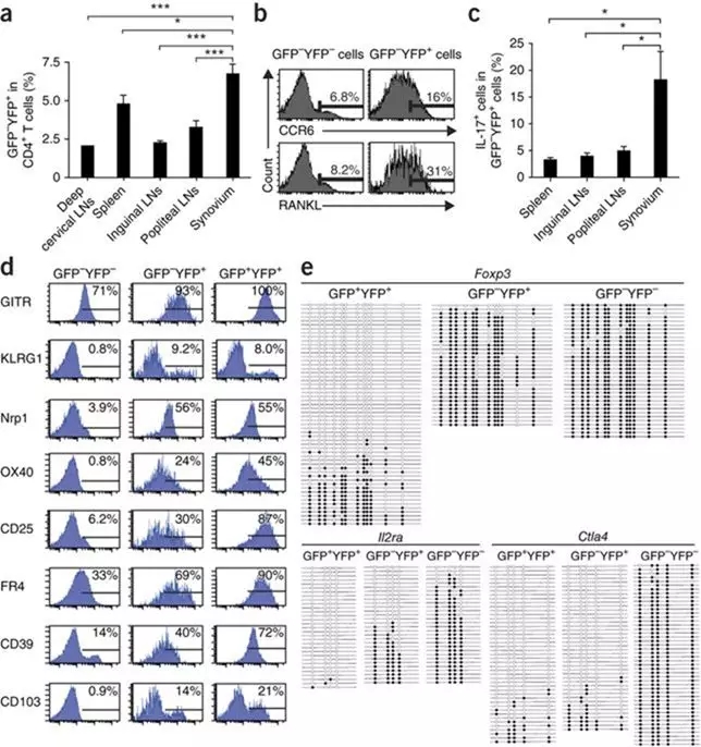
Figure 2 Localization, marker gene expression and DNA methylation status of exFoxp3 T cells in arthritic mice. Results are shown from fate mapping analyses of arthritic Foxp3-GFP-Cre × ROSA26-YFP mice that were performed 2 weeks after secondary immunization. (a) Frequency of exFoxp3 (GFP−YFP+) cells in CD4+ T cells isolated from the indicated location (n = 3 for deep cervical LNs, n = 8 for all other groups). (b) Expression of CCR6 and RANKL in the indicated T cell populations in popliteal lymph nodes. Representative data of six independent experiments are shown. (c) Frequency of IL-17+ cells in the GFP−YFP+ cell population (n = 6). (d) Expression of multiple Treg cell phenotypic markers in exFoxp3 T cells. Representative data of six independent experiments are shown. The percentages in each plot show the frequency of positive cells (indicated by the horizontal lines). (e) CpG methylation of the Foxp3, Il2ra and Ctla4 loci in exFoxp3 (GFP−YFP+), GFP+YFP+ and GFP−YFP−CD4+ T cells. A horizontal row within each box corresponds to one sequenced clone in which specific CpGs were methylated (closed) or demethylated (open). All data are shown as the mean ± s.e.m. Statistical analyses were performed using unpaired two-tailed Student’s t test (*P < 0.05, ***P < 0.005).
第三问:怎么成为我?
(小编:等等等等,不按逻辑出牌呢,不是应该问我要去哪么;我:去去,别打岔,我有更深刻的问题要思考,我是怎么成为现在这个样子的?这对理解关节炎发生的机制很重要)
上述实验结果中可以发现ex Foxp3 TH17细胞在炎症关节中聚集,作者假设关节中存在细胞与Foxp3+T细胞反应,促使其转变为TH17细胞。设计实验从关节炎小鼠中分离Thy1+CD11b-细胞(滑膜成纤维细胞)和Thy1-CD11b+细胞(滑膜巨噬细胞)与Foxp3+CD4+T细胞(从未处理的Foxp3hCD2小鼠中分离)共同培养, Thy1+CD11b-细胞下调了Foxp3+CD4+T细胞中Foxp3表达(图3a)。将 Thy1+CD11b-细胞与Foxp3+CD4+T细胞或naïve CD4+ T细胞共同培养,检测IL17+Foxp3-细胞比例变化及细胞因子的表达(IFN-γ、IL-4),结果显示滑膜成纤维细胞使exFoxp3 T细胞上调了IL-17的表达水平,IFN-γ和IL-4表达没有变化,表明滑膜成纤维细胞在关节中促使Foxp3+T细胞转变为TH17细胞。Naïve CD4+ T细胞没有促使发生这一转变。进一步研究这一转变发生的机制,将Thy1+CD11b-细胞和与Foxp3+CD4+T细胞共同培养的上清液与细胞分离分别检测(图3d),发现细胞并不能引起这个转变;检测和分析上清液的细胞因子表达,结果显示Thy+CD11b-细胞上清产生较高的IL-6因子,并在IL-17存在的情况下,IL-6表达进一步加强(图e)。加入抗IL-6抗体抑制了ex Foxp3+ TH17细胞的产生。证明来源于滑膜成纤维细胞的IL-6是促使Foxp3+CD4+T细胞转变为TH17细胞的关键因素。
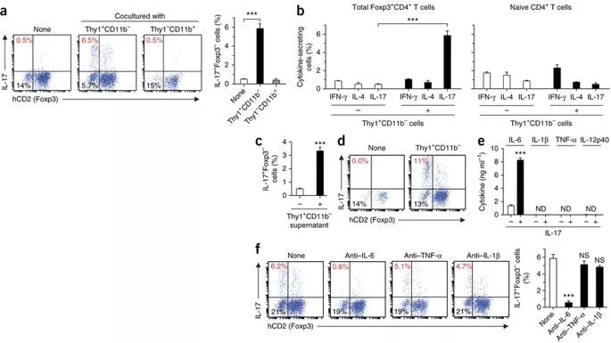
Figure 3 Arthritic synovial fibroblasts promote the conversion of Foxp3+ T cells to TH17 cells in an IL-6– dependent manner. (a) Coculture of Foxp3+CD4+ T cells and Thy1+CD11b− synovial fibroblasts or Thy1−CD11b+ synovial macrophages isolated from arthritic mice. The frequencies of hCD2+ (Foxp3) and IL-17+ cells were examined after 3 d of coculture. Representative data (left) and quantification (n = 3; right) are shown. (b) IFN-γ, IL-4 and IL-17 expression (n = 3 per analysis) in cocultures of total Foxp3+CD4+ T cells (left) or naive CD4+ T cells (right) with Thy1+CD11b− synovial fibroblasts. (c,d) Frequency of IL-17+Foxp3− cells after 3 d of culture of total Foxp3+CD4+ T cells with the supernatant of Thy1+CD11b− synovial fibroblasts (c; n = 3) or with Thy1+CD11b− synovial fibroblasts using a transwell chamber (d; representative data). (e) Cytokine production by Thy1+CD11b− synovial fibroblasts cultured in the presence or absence of IL-17 (n = 3). (f) Left, expression of Foxp3 and IL-17 in T cells after culture of Foxp3+CD4+ T cells with Thy1+CD11b− synovial fibroblasts in the presence of neutralizing antibodies to IL-6, TNF-α or IL-1β (representative data). Right, quantitative analysis of the frequency of IL-17+Foxp3− cells in T cells (n = 3). All data are shown as the mean ± s.e.m. Statistical analyses were performed using unpaired two-tailed Student’s t test (***P < 0.005; NS, not significant; ND, not detected). Data are representative of three independent experiments with triplicate culture wells.
第四问:我有什么高(xie)超(e)的能力?-促进破骨细胞生成
破骨细胞是造成关节炎骨破坏的重要因素。为了评估TH17细胞在关节炎中对骨破坏的影响程度,作者通过破骨细胞(TRAP染色的多核细胞,TRAP+MNs)计数和RANKL、细胞因子表达,检测其诱导破骨细胞生成的能力。将Foxp3hCD2小鼠与IL-17-GFP小鼠(该小鼠来源于百奥赛图哦)交配,方便提取IL-17表达细胞。exFoxp3 TH17细胞、Foxp3+T细胞、IL17a-/-TH17细胞分别与滑膜成纤维细胞、骨髓源肥大和巨噬前体细胞(BMMs)共同培养,结果显示,ex Foxp3 TH17细胞比TH17细胞(naïve CD4+T细胞来源的)具有更强的破骨细胞生成能力(图4a,b)。 TH17细胞的诱导破骨细胞生成能力主要来自IL-17的表达,IL-17可以刺激成纤维细胞上RANKL表达, TH17细胞虽然本身也产生RANKL,但细胞自身单独没有办法诱导破骨细胞生成。而IL17a-/-ex Foxp3 TH17细胞缺失IL-17A表达仍可以诱导破骨细胞生成,说明是T细胞来源的RANKL或细胞因子而不是IL-17A对破骨细胞生成有潜在影响(图4b)。作者发现ex Foxp3 TH17细胞比naïve CD4+T细胞来源的TH17细胞 表达了更高水平的RANKL,经过滑膜成纤维细胞共同培养,RANKL表达进一步增强。(图4c)
如图4d,ex Foxp3 TH17细胞与BMMs共同培养,发现在没有滑膜成纤维细胞存在的时候, ex Foxp3 TH17细胞也有将BMMs诱导生成破骨细胞的能力。为了探究RANKL对ex Foxp3 TH17细胞和滑膜成纤维细胞的影响,文中在共同培养系统中使用ex Foxp3 TH17细胞(从Lck-cre(T淋巴细胞特异cre小鼠) Tnfsf11(编码RANKL)flox/△ Foxp3hCD2 小鼠中分离)或Tnfsf11缺失的滑膜成纤维细胞。结果表示RANKL的表达是滑膜成纤维细胞促使破骨细胞生成的关键因素,而在没有表达RANKL的滑膜成纤维细胞存在下,ex Foxp3 TH17细胞 中RANKL的表达也能够促使破骨细胞生成(图4e,f)。
用GeneChip分析ex Foxp3 TH17细胞和TH17细胞(naïve CD4+T细胞来源的), 相比之下,ex Foxp3 TH17细胞表达了更高的Ccr6、Ccl20、Il23r,Il17re,Tnfrsf21,Vcam1,Rorc和Tnfsf11,较低的Csf2,Tnf,Il10和Sgk1。结果说明ex Foxp3 TH17细胞与多数已知的致病性的TH17细胞亚群并不相同,组成一种新的致病性TH17细胞亚群。
看看,同样都是TH17细胞,我是ex Foxp3 TH17细胞,我的祖先是exFoxp3 T细胞,通过获得了激活TH17变化而来的,跟其他TH17细胞就是不一样哦! (高唱:“我们不一样!”)
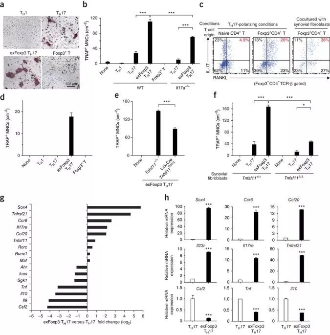
Figure 4 exFoxp3 TH17 cells are osteoclastogenic T cells with distinct gene profiles. (a,b) Osteoclast differentiation in a coculture of BMMs, Thy1+CD11b− CIA synovial fibroblasts and the T cell subsets indicated. exFoxp3 TH17 indicates Foxp3− T cells developed from Foxp3+ T cells under TH17-polarizing conditions in this experiment. (a) Representative tartrate-resistant acid phosphatase (TRAP) staining. (b) Number of osteoclasts (TRAP+ MNCs) (n = 3 for exFoxp3 TH17 and Foxp3+ T cells, n = 4 for Il17a−/− TH17 cells, n = 6 for all other groups). WT, wild type. (c) Representative FACS profiles of RANKL and IL-17 expression in naive CD4+ T cell–derived TH17 cells (left) and exFoxp3 TH17 cells (middle and right) that differentiated under the conditions indicated. (d) Number of osteoclasts (TRAP+ MNCs) in a coculture of BMMs and the T cell subsets indicated (n = 3). (e,f) Number of osteoclasts (TRAP+ MNCs) in a coculture of BMMs, synovial fibroblasts and the T cell subsets indicated. (e) Analysis using exFoxp3 TH17 cells derived from Tnfsf11+/+ Foxp3hCD2 or Lck-Cre Tnfsf11flox/Δ Foxp3hCD2 mice. (f) Analysis using Tnfsf11+/+ or Tnfsf11Δ/Δ arthritic synovial fibroblasts. (g) Microarray analysis of selected TH17-related genes in exFoxp3 TH17 cells and TH17 cells. IL-17GFP+ Foxp3hCD2− cells developed from naive CD4+ T cells or Foxp3+CD4+ T cells under TH17-polarizing conditions were used as the TH17 cells and exFoxp3 TH17 cells, respectively. The mean fold change of three independent experiments is shown. (h) Quantitative RT-PCR analysis of differentially expressed genes in TH17 cells and exFoxp3 TH17 cells (n = 3). All data are representative of three independent experiments with triplicate culture wells and are shown as the mean ± s.e.m. Statistical analyses were performed using unpaired two-tailed Student’s t test (*P < 0.05, ***P < 0.005).
第五问:我的家族在关节炎中有哪些贡献?
我的祖先是ex Foxp3 T细胞,来自CD25loFoxp3+ CD4+ T细胞家族,我们家族好像很厉害的样子呢,让我们一起来看一看它们对关节炎有哪些“贡献”吧。
胸腺来源的稳定的Foxp3+CD4+ T细胞与自身抗原有较高的亲和力,维持了自身耐受性。作者假设不稳定的Foxp3+CD4+T细胞也包含自我反应T细胞,并且在失去Foxp3表达之后导致关节炎发生。为了研究自身抗原特异性的exFoxp3 T细胞的作用,作者将CD25loFoxp3+、 CD25hiFoxp3+、Foxp3-、效应记忆CD44hiFoxp3-CD4+T细胞(来源于胶原免疫的DBA/1 Foxp3hCD2小鼠)标记CFSE,转移到免疫小鼠模型中, CD25loFoxp3+细胞加速了关节炎发生,加重了症状。关节炎条件下,自身反应CD4+ T 细胞主要来源于CD25loFoxp3+ CD4+ T细胞(图5a-c)。相比, CD25hiFoxp3+细胞显著抑制了破骨细胞的生成,几乎一半的CD25loFoxp3+细胞失去了Foxp3表达,几乎全部CD25hiFoxp3+细胞仍表达Foxp3(图5d)。体外二型胶原刺激后, CD25loFoxp3+细胞开始增殖,表明其包含更多的自身反应T细胞(图5e)。

三、场景再现
场景:关节炎
主人公: ex Foxp3 TH17细胞
我是ex Foxp3 TH17细胞,来自pTreg细胞部落,从CD25loFoxp3+ CD4+ T细胞丢失Foxp3表达,聚集到关节,经滑膜成纤维细胞产生的IL-6转变而来。表达Ccr6、CCL20、IL23R、RANKL是我的标志,有较强的促破骨细胞发生能力,在关节炎的病理中有比较关键的作用。希望通过我的独白大家能够了解自身免疫关节炎中的发生机制和关键角色,我也能成为RA的标志物,预测抗IL-6抗体的治疗效果,进而发展成为自身免疫疾病的新的治疗手段。
本篇文章作者充分利用了基因编辑小鼠研究了自身免疫疾病的部分发生机制。很荣幸百奥赛图起家第一桶金的产品鼠的IL-17a GFP KI小鼠对文章的研究有所帮助,如果您需要购买该小鼠或制备属于您的专属科研小鼠,请与我们联系哦!
参考文献:
1. Komatsu, N., Okamoto, K., Sawa, S., Nakashima, T., Oh-hora, M., Kodama, T., … Takayanagi, H. (2013). Pathogenic conversion of Foxp3+ T cells into TH17 cells in autoimmune arthritis. Nature Medicine, 20(1), 62–68.
2. Zhou, X., Jeker, L. T., Fife, B. T., Zhu, S., Anderson, M. S., McManus, M. T., & Bluestone, J. A. (2008). Selective miRNA disruption in T reg cells leads to uncontrolled autoimmunity. The Journal of Experimental Medicine, 205(9), 1983–1991.
3. Zhou, X., Bailey-Bucktrout, S. L., Jeker, L. T., Penaranda, C., Martínez-Llordella, M., Ashby, M., … Bluestone, J. A. (2009). Instability of the transcription factor Foxp3 leads to the generation of pathogenic memory T cells in vivo. Nature Immunology, 10(9), 1000–1007.