
单细胞应力加载测试分析系统
产品名称: 单细胞应力加载测试分析系统
英文名称: Single cell stress loading analysis system
产品编号: Optical Stretcher
产品价格: 0
产品产地: 德国
品牌商标: rszelltechnik
更新时间: null
使用范围: null
- 联系人 :
- 地址 : 北京市丰台区马家堡西路36号东亚·三环1号写字楼171
- 邮编 :
- 所在区域 : 北京
- 电话 : 186****2672
- 传真 : 010-57129142
- 邮箱 : shengliangli@bio-goods.com
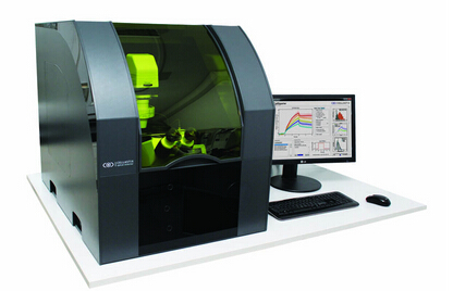
单细胞高通量细胞激光牵张拉伸应力加载与力学属性分析系统
Optical Stretcher是用于细胞生物力学高通量测量研究的激光光学牵张拉伸创新平台技术。
世界第一台用来高通量测量单个悬浮细胞(悬液细胞)的变形能力设备。
该激光光学牵张拉伸器是个可以安装在任何相位差显微镜上的模块。温度稳定和激光安全的显微镜系统。
Optical Stretcher 
创新研究(Innovation in Research)
Optical Stretcher激光光学牵张拉伸器是一种新颖的用来测量和分析单个悬液细胞生物力学特性(比如:如弹性和松弛)的激光工具。
非接触式细胞形变(Contact-free cell deformation)
无接触式细胞形变“开放=”0“的风格=”2“]是激光力引起的悬浮细胞形变,这决定绝对的无接触式测量。这可确保均匀的细胞处理,避免因接触引起的细胞反应文物。
高通量单细胞流变
通过集成的微流体系统可以很可以容易地测定300个细胞/小时。这样就可以在第一时间收集细胞流变显著的统计数据
省时,自动测量(Timesaving, automated measurements)
对应于用户定义的拉伸模式,细胞被自动传送到测量区域进行形变。在光学拉伸加载运行实验中,你可以专注于阐述实验结果。
产品规格
-
包括有两个压力控制通道的微流体系统
-
最大每个光纤2功率W掺镱光纤激光器
-
安装倒置相差显微镜
-
激光安全和温度控制
-
可选用组合荧光显微镜
软件规格
-
使用CellStretcher模块控制所有组件和自动测量细胞
-
由CellEvaluator提取记录显微图像形变数据
-
由CellReporter统计分析和可视化特性参数
-
为自己的统计分析访问原始数据
产品特点:
-
非接触式和无标记的细胞测量
-
高通量-250细胞/小时
-
省时的自动测量
-
模块可以在任何倒置相差显微镜进行安装
-
外壳激光安全和稳定的温度
-
数据评估软件
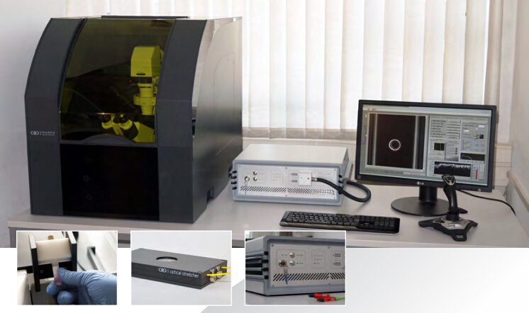
产品规格:
Fibolux laser system 2 W
reliable microfluidic system for easy probe handling
尺寸 cm (w x h x d): 70 x 80 x 100
Options
combination with fluorescence microscopy

技术
optical stretcher 是一种新颖的微操纵单个生物细胞激光工具,探讨在悬浮液的粘弹性性质[1]。


通过两个对立的激光束钳持一个细胞,进行牵张拉伸细胞两边。更高的激光功率使细胞发生形变。
细胞的形变是由CCD相机记录,并由一专门设计的软件进行评估。
Optical Stretcher 测量室集成有微流系统,使得细胞容易地一个接一个地输送
可以达到每小时约250个细胞的高吞吐率,允许相对于其它工具,例如原子力显微镜(AFM)更好的统计信息。


Momentum transfer at the cell surface
Cell mechanics as a disease marker
The physical mechanics of cells are important for their regular, biological functioning and are regulated by a structure called the cytoskeleton. It is involved in many vital processes of the cell. If these are changes this naturally results also in changes of the biomechanical properties, which can be measured with the Optical Stretcher. There is already published data for cancer [2, 3] and for the effect of cell aging [4].
Several ongoing studies examine the ability of the Optical Stretcher to differentiate between the stages of a cancer tumor, making it a valuable tool for both scientific research and clinical diagnosis [5].

Cell types can be differentiated by their deformation in the optical stretcher
细胞形变力源自激光。当光被折射在细胞表面存在的光子的动量的变化。因为整体动量必须始终保守有一个在垂直于作用于它的力形式的动量转移到细胞表面。
Research
Our device allows various applications in basic research of biophysics, biology & medicine.
Industry
Due to the high-throughput the Optical Stretcher is suitable for industrial drug-screening.
Clinical Diagnostic
First clinical trials with breast cancer tumors show a different deformation of cancerous cells.

应用:
生物物理研究(Biophysical Research)
Characterization of fundamental cytoskeletal functions and processes in eukaryotic cells
药物筛选(Drug Screening)
Testing new substances and their efficiency on a cellular basis
Aging proscesses
Identification of markers for cell aging and testing of anti-aging substances
干细胞分化(Differentiation)
Utilization of cell stiffness as a marker for differentiation processes in stem cells
Immune response
Investigation of cytoskeletal changes of immune-activated cells
Mechanisms of diseases
New insight in cellular changes caused by diseases such as cancer, malaria or sepsis
Publications
Optical Stretcher Technology
Lincoln, B., Schinkinger, S., Travis, K., Wottawah, F., Ebert, S., Sauer, F., Guck, J., 2007. Reconfigurable microfluidic integration of a dual-beam laser trap with biomedical applications. Biomed. Microdevices 9, 703–710. doi:10.1007/s10544-007-9079-x
Ebert, S., Travis, K., Lincoln, B., Guck, J., 2007. Fluorescence ratio thermometry in a microfluidic dual-beam laser trap.Opt. Express 15, 15493–15499. doi:10.1364/OE.15.015493
Jensen-McMullin, C., Lee, H.P., Lyons, E.R.L., 2005. Demonstration of trapping, motion control, sensing and fluorescence detection of polystyrene beads in a multi-fiber optical trap. Opt. Express 13, 2634–2642. doi:10.1364/OPEX.13.002634
Wottawah, F., Schinkinger, S., Lincoln, B., Ananthakrishnan, R., Romeyke, M., Guck, J., K?s, J., 2005.Optical Rheology of Biological Cells. Phys. Rev. Lett. 94, 098103. doi:10.1103/PhysRevLett.94.098103
Lincoln, B., Erickson, H.M., Schinkinger, S., Wottawah, F., Mitchell, D., Ulvick, S., Bilby, C., Guck, J., 2004. Deformability-based flow cytometry.Cytometry A 59A, 203–209. doi:10.1002/cyto.a.20050
Theoretical Models
Ananthakrishnan, R., Guck, J., Wottawah, F., Schinkinger, S., Lincoln, B., Romeyke, M., Kas, J., 2005. Modelling the structural response of an eukaryotic cell in the optical stretcher. Curr. Sci. 88.
B. Bareil, P., Sheng, Y., Chiou, A., 2006. Local scattering stress distribution on surface of a spherical cell in optical stretcher. Opt. Express 14, 12503–12509. doi:10.1364/OE.14.012503
Bareil, P.B., Sheng, Y., Chen, Y.-Q., Chiou, A., 2007. Calculation of spherical red blood cell deformation in a dual-beam optical stretcher. Opt. Express 15, 16029–16034. doi:10.1364/OE.15.016029
Boyde, L., Ekpenyong, A., Whyte, G., Guck, J., 2012. Comparison of stresses on homogeneous spheroids in the optical stretcher computed with geometrical optics and generalized Lorenz–Mie theory. Appl. Opt. 51, 7934–7944. doi:10.1364/AO.51.007934
Ekpenyong, A.E., Posey, C.L., Chaput, J.L., Burkart, A.K., Marquardt, M.M., Smith, T.J., Nichols, M.G., 2009. Determination of cell elasticity through hybrid ray optics and continuum mechanics modeling of cell deformation in the optical stretcher.Appl. Opt. 48, 6344–6354. doi:10.1364/AO.48.006344
Teo, S.-K., Goryachev, A.B., Parker, K.H., Chiam, K.-H., 2010. Cellular deformation and intracellular stress propagation during optical stretching.Phys. Rev. E 81, 051924. doi:10.1103/PhysRevE.81.051924
Cancer research and diagnostics
Martin, M., Müller, K., Cadenas, C., Hermes, M., Zink, M., Hengstler, J.G., K?s, J.A., 2012. ERBB2 overexpression triggers transient high mechanoactivity of breast tumor cells. Cytoskeleton 69, 267–277. doi:10.1002/cm.21023
Fritsch, A., H?ckel, M., Kiessling, T., Nnetu, K.D., Wetzel, F., Zink, M., K?s, J.A., 2010. Are biomechanical changes necessary for tumour progression?Nat. Phys. 6, 730–732. doi:10.1038/nphys1800
Brunner, C., Niendorf, A., K?s, J.A., 2009. Passive and active single-cell biomechanics: a new perspective in cancer diagnosis. Soft Matter 5, 2171–2178. doi:10.1039/B807545J
Remmerbach, T.W., Wottawah, F., Dietrich, J., Lincoln, B., Wittekind, C., Guck, J., 2009. Oral Cancer Diagnosis by Mechanical Phenotyping. Cancer Res. 69, 1728–1732. doi:10.1158/0008-5472.CAN-08-4073
Martin, M., Mueller, K., Wottawah, F., Schinkinger, S., Lincoln, B., Romeyke, M., K?s, J.A., 2006. Feeling with light for cancer. p. 60800P–60800P–10. doi:10.1117/12.637899
Guck, J., Schinkinger, S., Lincoln, B., Wottawah, F., Ebert, S., Romeyke, M., Lenz, D., Erickson, H.M., Ananthakrishnan, R., Mitchell, D., K?s, J., Ulvick, S., Bilby, C., 2005. Optical Deformability as an Inherent Cell Marker for Testing Malignant Transformation and Metastatic Competence. Biophys. J. 88, 3689–3698. doi:10.1529/biophysj.104.045476
Stem cell research
Ekpenyong, A.E., Whyte, G., Chalut, K., Pagliara, S., Lautenschlaeger, F., Fiddler, C., Paschke, S., Keyser, U.F., Chilvers, E.R., Guck, J., 2012.Viscoelastic Properties of Differentiating Blood Cells Are Fate- and Function-Dependent. Plos One 7, e45237. doi:10.1371/journal.pone.0045237
Galle, J., Bader, A., Hepp, P., Grill, W., Fuchs, B., Kas, J.A., Krinner, A., MarquaB, B., Muller, K., Schiller, J., Schulz, R.M., von Buttlar, M., von der Burg, E., Zscharnack, M., Loffler, M., 2010. Mesenchymal Stem Cells in Cartilage Repair: State of the Art and Methods to monitor Cell Growth, Differentiation and Cartilage Regeneration. Curr. Med. Chem. 17, 2274–2291. doi:10.2174/092986710791331095
Maloney, J.M., Nikova, D., Lautenschlager, F., Clarke, E., Langer, R., Guck, J., Van Vliet, K.J., 2010. Mesenchymal Stem Cell Mechanics from the Attached to the Suspended State. Biophys. J. 99, 2479–2487. doi:10.1016/j.bpj.2010.08.052
Lautenschl?ger, F., Paschke, S., Schinkinger, S., Bruel, A., Beil, M., Guck, J., 2009. The regulatory role of cell mechanics for migration of differentiating myeloid cells. Proc. Natl. Acad. Sci. 106, 15696–15701. doi:10.1073/pnas.0811261106
Basic research
Gyger, M., Stange, R., Kiessling, T.R., Fritsch, A., Kostelnik, K.B., Beck-Sickinger, A.G., Zink, M., Kaes, J.A., 2014. Active contractions in single suspended epithelial cells. Eur. Biophys. J. Biophys. Lett. 43, 11–23. doi:10.1007/s00249-013-0935-8
Seltmann, K., Fritsch, A.W., K?s, J.A., Magin, T.M., 2013. Keratins significantly contribute to cell stiffness and impact invasive behavior. Proc. Natl. Acad. Sci. 201310493. doi:10.1073/pnas.1310493110
Kie?ling, T.R., Stange, R., K?s, J.A., Fritsch, A.W., 2013. Thermorheology of living cells—impact of temperature variations on cell mechanics. New J. Phys. 15, 045026. doi:10.1088/1367-2630/15/4/045026
Kie?ling, T.R., Herrera, M., Nnetu, K.D., Balzer, E.M., Girvan, M., Fritsch, A.W., Martin, S.S., K?s, J.A., Losert, W., 2013. Analysis of multiple physical parameters for mechanical phenotyping of living cells. Eur. Biophys. J. 42, 383–394. doi:10.1007/s00249-013-0888-y
Paschke, S., Weidner, A.F., Paust, T., Marti, O., Beil, M., Ben-Chetrit, E., 2013. Technical advance: Inhibition of neutrophil chemotaxis by colchicine is modulated through viscoelastic properties of subcellular compartments. J. Leukoc. Biol. 94, 1091–1096. doi:10.1189/jlb.1012510
Chalut, K.J., H?pfler, M., Lautenschl?ger, F., Boyde, L., Chan, C.J., Ekpenyong, A., Martinez-Arias, A., Guck, J., 2012. Chromatin decondensation and nuclear softening accompany Nanog downregulation in embryonic stem cells. Biophys. J. 103, 2060–2070. doi:10.1016/j.bpj.2012.10.015
Matthews, H.K., Delabre, U., Rohn, J.L., Guck, J., Kunda, P., Baum, B., 2012. Changes in Ect2 localization couple actomyosin-dependent cell shape changes to mitotic progression. Dev. Cell 23, 371–383. doi:10.1016/j.devcel.2012.06.003
Mauritz, J.M.A., Esposito, A., Tiffert, T., Skepper, J.N., Warley, A., Yoon, Y.-Z., Cicuta, P., Lew, V.L., Guck, J.R., Kaminski, C.F., 2010. Biophotonic techniques for the study of malaria-infected red blood cells. Med. Biol. Eng. Comput. 48, 1055–1063. doi:10.1007/s11517-010-0668-0
Rusciano, G., 2010. Experimental analysis of Hb oxy–deoxy transition in single optically stretched red blood cells. Phys. Med. 26, 233–239. doi:10.1016/j.ejmp.2010.02.001
Aging processes
Schulze, C., Wetzel, F., Kueper, T., Malsen, A., Muhr, G., Jaspers, S., Blatt, T., Wittern, K.-P., Wenck, H., K?s, J.A., 2010.Stiffening of Human Skin Fibroblasts with Age. Biophys. J. 99, 2434–2442. doi:10.1016/j.bpj.2010.08.026
Vesicles
Solmaz, M.E., Sankhagowit, S., Biswas, R., Mejia, C.A., Povinelli, M.L., Malmstadt, N., 2013. Optical stretching as a tool to investigate the mechanical properties of lipid bilayers. Rsc Adv. 3, 16632–16638. doi:10.1039/c3ra42510j
Solmaz, M.E., Biswas, R., Sankhagowit, S., Thompson, J.R., Mejia, C.A., Malmstadt, N., Povinelli, M.L., 2012. Optical stretching of giant unilamellar vesicles with an integrated dual-beam optical trap. Biomed. Opt. Express 3, 2419–2427. doi:10.1364/BOE.3.002419
Technical advances
Bellini, N., Bragheri, F., Cristiani, I., Guck, J., Osellame, R., Whyte, G., 2012. Validation and perspectives of a femtosecond laser fabricated monolithic optical stretcher. Biomed. Opt. Express 3, 2658–2668. doi:10.1364/BOE.3.002658
Bellini, N., Vishnubhatla, K.C., Bragheri, F., Ferrara, L., Minzioni, P., Ramponi, R., Cristiani, I., Osellame, R., 2010.Femtosecond laser fabricated monolithic chip for optical trapping and stretching of single cells. Opt. Express 18, 4679–4688. doi:10.1364/OE.18.004679