尼康 Nikon C2 Plus激光共聚焦显微镜
产品名称: 尼康 Nikon C2 Plus激光共聚焦显微镜
英文名称:
产品编号: C2 plus
产品价格: 0
产品产地: 日本
品牌商标: Nikon/尼康
更新时间: null
使用范围: null
An essential microscopy laboratory instrument
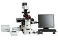
 Configured with Ni-E
The C2+ confocal microscope system is part of a new generation of Nikon confocal instruments designed to be essential laboratory microscopy tools. Built on a reputation of incredible stability and operational simplicity coupled with superior optical technologies and high-speed image acquisition of up to 100 fps*, the C2+ is the perfect tool for a new microscope, or as a new accessory to an existing Nikon imaging system.
- *With 8x zoom or larger
Large field-of-view imaging and three-dimensional reconstruction
Confocal image acquisition, using high-numerical aperture and high-magnification objectives, together with XY stage control and advanced image stitching with Nikon NIS-Elements software, enables high-resolution images of large areas of a specimen to be produced. In addition, the microscope's high-precision Z-axis control allows assemblage of Z stack images for three-dimensional image reconstruction.
Image Stitching (Large Image)

Specimen: Genital tract of Drosophila melanogaster
Photo courtesy of: Director and Professor Masatoshi Yamamoto, Drosophila Genetic Resource Center, Kyoto Institute of Technology
Image Quality
Nikon's unprecedented optics and a time-proven, highly efficient optical design provide the brightest and sharpest images, at the longest working distances.
High-efficiency scanning heads and detectors

With the convenient, small scan head size, the C2+ can be used with various types of Nikon microscope. The C2+ employs high precision mirrors and optically superior circular pinholes, and separates the detectors to isolate sources of heat and noise, enabling low-noise, high-contrast and high-quality confocal imaging. The newly developed scanner driving system and Nikon's unique image correction technique allow 8 fps (512 x 512 pixels) and 100 fps (512 x 32 pixels) high-speed imaging.
High-performance optics

CFI Apochromat 40xWI λS, NA1.25 (left)
CFI Apochromat LWD 40xWI λS, NA1.15 (middle)
CFI Apochromat 60x Oil λS, NA1.4 (right)
CFI Apochromat λS Series
These high-numerical aperture (NA) objectives are ideal for confocal imaging with correction of chromatic aberrations over a wide wavelength range from ultraviolet. In particular, the LWD 40xWI lens corrects up to infrared. Transmission is increased through the use of Nikon's exclusive Nano Crystal Coat technology.
CFI Apochromat TIRF Series

CFI Apochromat TIRF 60x oil, NA1.49 (left)
CFI Apochromat TIRF 100x oil, NA1.49 (right)
These objectives boast an unprecedented NA of 1.49 (using a standard coverslip and immersion oil), the highest resolution among Nikon objectives. The temperature correction ring corrects image quality affected by temperature change in the range of 23°C to 37°C.
High-definition diascopic DIC images
The C2+ can acquire simultaneous three-channel fluorescence or simultaneous three-channel and diascopic DIC observation. High-quality DIC images and fluorescence images can be superimposed to aid in morphological analysis.

DIC image

Overlay of DIC and fluorescence images
High functionality
High-performance imaging software NIS-Elements offers a variety of image processing and analysis functions. It also enables data extraction from acquired images. In addition, NIS-Elements allows for intuitive operation of Nikon microscopes and other third-party peripheral devices, such as EMCCD cameras and filter wheels, to broaden the range of experiments possible.
Multimode capability
Various imaging methods, such as confocal, widefield, TIRF, photoactivation, as well as processing, analysis and presentation of acquired images, are available in one software package. Users can easily learn how to control different imaging systems with a common interface and workflow.

Easy-to-recognize display for setting lasers, detectors, etc.

Scanning parameter settings
Unmixing

Spectral analysis GUI
Numerous functions for analysis and unmixing of acquired spectrums are provided, while spectral profiles of general dyes and fluorescent proteins are preprogrammed.
Flexibility
The C2+ can be coupled with upright, inverted, physiological, and macro imaging microscopes and has options for combinations with various high-quality research experiment systems. All can be controlled with NIS-Elements software.
TIRF/Photoactivation-C2+ Multimode imaging system

Optional TIRF laser illumination module and a photoactivation module can be integrated to enable both imaging of single molecules with an extremely high S/N ratio, and imaging of the fluorescence characteristic changes of photoactivated and photo-convertible fluorescent protein.
AZ-C2+ Macro confocal microscope system

With a high-definition large field of view, specimens larger than 1cm can be acquired with an unprecedentedly high S/N ratio. The AZ-C2+ allows for imaging of whole-mount specimens, such as embryos, in a single acquisition, up to 4000x4000 pixel resolution, and it can also acquire 32-channel spectral data with the C2si+. It offers a combination of low and high magnification objective lenses, optical zoom and a confocal scanning zoom function, enabling continuous imaging from macro to micro.

TT2 ES cells
Anti-Nanog antibody (Cy3), anti-Oct3/4 antibody (Alexa488) and DAPI localized in cell nuclei
Photographed with the cooperation of: Hiroshi Kiyonari, Laboratory for Animal Resources and Genetic Engineering, RIKEN Center for Developmental Biology

Photo courtesy of: Director and Professor Masatoshi Yamamoto, Drosophila Genetic Resource Center, Kyoto Institute of Technology
Some sample images in this brochure were captured using the C1 confocal microscope system.