NG2,黑色素瘤硫酸软骨素蛋白多糖4抗体
产品名称: NG2,黑色素瘤硫酸软骨素蛋白多糖4抗体
英文名称: Anti-NG2 antibody
产品编号: HZ-11192R
产品价格: null
产品产地: 中国/美国
品牌商标: HZbscience
更新时间: 2023-08-17T10:24:20
使用范围: WB,ELISA,IHC-P,IHC-F,IF
- 联系人 : 鲍丽雯
- 地址 : 上海市闵行区闵北路88弄1-30号第22幢AQ136室
- 邮编 : 200612
- 所在区域 : 上海
- 电话 : 139****0749
- 传真 : 021-60345367
- 邮箱 : www.shzbio.net
NG2,黑色素瘤硫酸软骨素蛋白多糖4抗体
产品编号HZ-11192R
英文名称NG2
中文名称黑色素瘤硫酸软骨素蛋白多糖4抗体
别 名MELCSPG; AN2; AN2 proteoglycan; Chondroitin sulfate proteoglycan 4 (melanoma-associated); Chondroitin sulfate proteoglycan 4; Chondroitin sulfate proteoglycan NG2; CSPG4; Cspg4 chondroitin sulfate proteoglycan 4; CSPG4_HUMAN; HMW-MAA; HSN tumor-specific antigen; Kiaa4232; MCSP; MCSPG; MEL-CSPG; Melanoma chondroitin sulfate proteoglycan; Melanoma-associated chondroitin sulfate proteoglycan; 4732461B14Rik; MSK16.
说 明 书0.1ml 0.2ml
研究领域肿瘤 细胞生物 免疫学 神经生物学
抗体来源Rabbit
克隆类型Polyclonal
交叉反应 Human, Mouse, Rat, Pig, Cow,
NG2,黑色素瘤硫酸软骨素蛋白多糖4抗体产品应用ELISA=1:500-1000 IHC-P=1:100-500 IHC-F=1:100-500 ICC=1:100-500 IF=1:100-500 (石蜡切片需做抗原修复)
not yet tested in other applications.
optimal dilutions/concentrations should be determined by the end user.
分 子 量247kDa
细胞定位细胞浆 细胞膜
性 状Lyophilized or Liquid
浓 度1mg/1ml
免 疫 原KLH conjugated synthetic peptide derived from human NG2
亚 型IgG
纯化方法affinity purified by Protein A
储 存 液0.01M TBS(pH7.4) with 1% BSA, 0.03% Proclin300 and 50% Glycerol.
保存条件Store at -20 °C for one year. Avoid repeated freeze/thaw cycles. The lyophilized antibody is stable at room temperature for at least one month and for greater than a year when kept at -20°C. When reconstituted in sterile pH 7.4 0.01M PBS or diluent of antibody the antibody is stable for at least two weeks at 2-4 °C.
NG2,黑色素瘤硫酸软骨素蛋白多糖4抗体PubMedPubMed
产品介绍background:
NG2 (also known as melanoma-associated chondroitin sulfate proteoglycan 4, MCSP, MCSPG, MSK16 and MEL-CSPG) stabilizes cell-substratum interactions during early events of melanoma cell spreading on endothelial basement membranes. NG2 may facilitate primary melanoma progression by enhancing the activation of key signaling pathways important for tumor invasion and growth. Threonine 2256 phosphorylation of rat NG2 (Threonine 2252 phosphorylation of human NG2) leads to redistribution of NG2 on the surface of astrocytomas, polarization of the cell and a significant increase in cell motility. NG2 acts as a co-receptor for spreading and focal contact formation in association with Alpha 4 Beta1 integrin in malignant melanoma cells. NG2 is present on blood vessels throughout the rat embryo. Microvessels within the rat CNS express NG2 on endothelial cells, and outside the CNS, NG2 is present on smooth muscle cells. NG2 is a novel marker for epidermal stem cells that contributes to their patterned distribution by promoting stem cell clustering.
Function:
Proteoglycan playing a role in cell proliferation and migration which stimulates endothelial cells motility during microvascular morphogenesis. May also inhibit neurite outgrowth and growth cone collapse during axon regeneration. Cell surface receptor for collagen alpha 2(VI) which may confer cells ability to migrate on that substrate. Binds through its extracellular N-terminus growth factors, extracellular matrix proteases modulating their activity. May regulate MPP16-dependent degradation and invasion of type I collagen participating in melanoma cells invasion properties. May modulate the plasminogen system by enhancing plasminogen activation and inhibiting angiostatin. Functions also as a signal transducing protein by binding through its cytoplasmic C-terminus scaffolding and signaling proteins. May promote retraction fiber formation and cell polarization through Rho GTPase activation. May stimulate alpha-4, beta-1 integrin-mediated adhesion and spreading by recruiting and activating a signaling cascade through CDC42, ACK1 and BCAR1. May activate FAK and ERK1/ERK2 signaling cascades.
Subunit:
Interacts with the first PDZ domain of MPDZ. Interacts with PRKCA. Binds TNC, laminin-1, COL5A1 and COL6A2. Interacts with PLG and angiostatin. Binds FGF2 and PDGFA. Interacts with GRIP1, GRIP2 and GRIA2. Forms a ternary complex with GRIP1 and GRIA2 (By similarity). Interacts with LGALS3 and the integrin composed of ITGB1 and ITGA3. Interacts with ITGA4 through its chondroitin sulfate glycosaminoglycan. Interacts with BCAR1, CDC42 and ACK1. Interacts with MMP16.
NG2,黑色素瘤硫酸软骨素蛋白多糖4抗体Subcellular Location:
Apical cell membrane. Cell projection > lamellipodium membrane. Localized at the apical plasma membrane it relocalizes to the lamellipodia of astrocytoma upon phosphorylation by PRKCA. Localizes to the retraction fibers. Localizes to the plasma membrane of oligodendrocytes.
Tissue Specificity:
Detected only in malignant melanoma cells.
Post-translational modifications:
O-glycosylated; contains glycosaminoglycan chondroitin sulfate which are required for proper localization and function in stress fiber formation (By similarity). Involved in interaction with MMP16 and ITGA4.
Phosphorylation by PRKCA regulates its subcellular location and function in cell motility (By similarity).
Similarity:
Contains 15 CSPG (NG2) repeats.
Contains 2 laminin G-like domains.
Gene ID:
1464
Database links:
Entrez Gene: 1464 Human
Entrez Gene: 121021 Mouse
Omim: 601172 Human
SwissProt: Q6UVK1 Human
SwissProt: Q8VHY0 Mouse
Unigene: 513044 Human
Unigene: 41329 Mouse
NG2,黑色素瘤硫酸软骨素蛋白多糖4抗体Important Note:
This product as supplied is intended for research use only, not for use in human, therapeutic or diagnostic applications.
| 产品图片 | 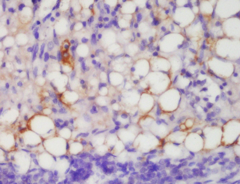
Tissue/cell: mouse melanoma tissue; 4% Paraformaldehyde-fixed and paraffin-embedded;
Antigen retrieval: citrate buffer ( 0.01M, pH 6.0 ), Boiling bathing for 15min; Block endogenous peroxidase by 3% Hydrogen peroxide for 30min; Blocking buffer (normal goat serum,C-0005) at 37℃ for 20 min; Incubation: Anti-NG2 Polyclonal Antibody, Unconjugated(HZ-11192R) 1:200, overnight at 4°C, followed by conjugation to the secondary antibody(SP-0023) and DAB(C-0010) staining 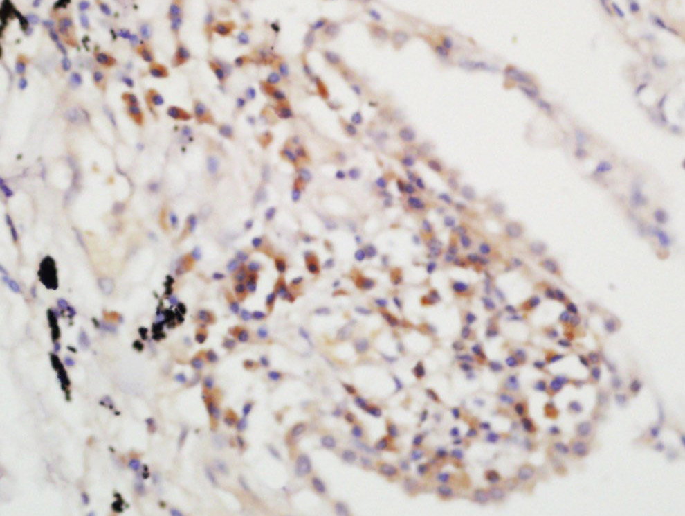
Tissue/cell: mouse lung carcinoma; 4% Paraformaldehyde-fixed and paraffin-embedded;
Antigen retrieval: citrate buffer ( 0.01M, pH 6.0 ), Boiling bathing for 15min; Block endogenous peroxidase by 3% Hydrogen peroxide for 30min; Blocking buffer (normal goat serum,C-0005) at 37℃ for 20 min; Incubation: Anti-NG2 Polyclonal Antibody, Unconjugated(HZ-11192R) 1:200, overnight at 4°C, followed by conjugation to the secondary antibody(SP-0023) and DAB(C-0010) staining |