Anti-TACD2抗体-抗体-抗体-生物在线
Anti-TACD2抗体

Anti-TACD2抗体

商家询价

产品名称: Anti-TACD2抗体

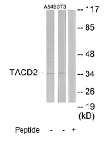
英文名称: Cell surface glycoprotein Trop 2 antibody Cell surface glycoprotein Trop-2 antibody Cell surface glycoprotein Trop2 antibody Epithelial glycoprotein 1

产品编号: ab65006

产品价格: null

产品产地: 英国

品牌商标: abcam

更新时间: null

使用范围:

深圳市宇德立生物科技有限公司
  • 联系人 :
  • 地址 : 深圳市宝安区西乡宝民二路贤基大厦4E
  • 邮编 :
  • 所在区域 : 广东
  • 电话 : 133****4454
  • 传真 : 0755-26457513
  • 邮箱 : 1484332550@qq.com


  • 形式Liquid
  • 存放说明Shipped at 4°C. Store at -20°C. Stable for 12 months at -20°C.
  • 存储溶液Preservative: 0.02% Sodium Azide
    Constituents: 50% Glycerol, PBS, 150mM Sodium chloride, pH 7.4
  • 浓度100 µg 浓度为 1 mg/ml 
  • 纯度Immunogen affinity purified
  • 纯化说明The antibody was affinity-purified from rabbit antiserum by affinity-chromatography using epitope-specific immunogen.
  • 克隆多克隆
  • 同种型IgG
  • 研究领域

应用

Our Abpromise guarantee covers the use of ab65006 in the following tested applications.

The application notes include recommended starting dilutions; optimal dilutions/concentrations should be determined by the end user.

应用 Abreviews 说明
IHC-P   Use a concentration of 5 µg/ml. Perform heat mediated antigen retrieval with citrate buffer pH 6 before commencing with IHC staining protocol.
WB   1/500 - 1/1000. Detects a band of approximately 37 kDa (predicted molecular weight: 37 kDa).
ELISA   1/5000.
ICC   Use at an assay dependent concentration.

靶标

  • 功能May function as a growth factor receptor.
  • 组织特异性Placenta, pancreatic carcinoma cell lines.
  • 疾病相关Defects in TACSTD2 are the cause of gelatinous drop-like corneal dystrophy (GDLD) [MIM:204870]; also known as lattice corneal dystrophy type III. GDLD is an autosomal recessive disorder characterized by grayish corneal amyloid deposits that cause severe visual impairment.
  • 序列相似性Belongs to the EPCAM family.
    Contains 1 thyroglobulin type-1 domain.
  • 翻译后修饰The N-terminus is blocked.
  • 细胞定位Membrane.
  • Anti-TACD2 antibody 图像

    • All lanes : Anti-TACD2 antibody (ab65006) at 1/500 dilution

      Lane 1 : Extracts from A549 cells
      Lane 2 : Extracts from NIH-3T3 cells
      Lane 3 : Extracts from NIH-3T3 cells with immunising peptide

      Lysates/proteins at 5 µg per lane.


      Predicted band size : 37 kDa
      Observed band size : 37 kDa
    • IHC image of ab65006 staining in human placenta formalin fixed paraffin embedded tissue section, performed on a Leica BondTM system using the standard protocol F. The section was pre-treated using heat mediated antigen retrieval with sodium citrate buffer (pH6, epitope retrieval solution 1) for 20 mins. The section was then incubated with ab65006, 5µg/ml, for 15 mins at room temperature and detected using an HRP conjugated compact polymer system. DAB was used as the chromogen. The section was then counterstained with haematoxylin and mounted with DPX.

      For other IHC staining systems (automated and non-automated) customers should optimize variable parameters such as antigen retrieval conditions, primary antibody concentration and antibody incubation times.